-
PDF
- Split View
-
Views
-
Cite
Cite
Kristian Thygesen, Joseph S. Alpert, Harvey D. White, Allan S. Jaffe, Fred S. Apple, Marcello Galvani, Hugo A. Katus, L. Kristin Newby, Jan Ravkilde, Bernard Chaitman, Peter M. Clemmensen, Mikael Dellborg, Hanoch Hod, Pekka Porela, Richard Underwood, Jeroen J. Bax, George A. Beller, Robert Bonow, Ernst E. Van Der Wall, Jean-Pierre Bassand, William Wijns, T. Bruce Ferguson, Philippe G. Steg, Barry F. Uretsky, David O. Williams, Paul W. Armstrong, Elliott M. Antman, Keith A. Fox, Christian W. Hamm, E. Magnus Ohman, Maarten L. Simoons, Philip A. Poole-Wilson, Enrique P. Gurfinkel, José-Luis Lopez-Sendon, Prem Pais, Shanti Mendis, Jun-Ren Zhu, Lars C. Wallentin, Francisco Fernández-Avilés, Kim M. Fox, Alexander N. Parkhomenko, Silvia G. Priori, Michal Tendera, Liisa-Maria Voipio-Pulkki, Alec Vahanian, A. John Camm, Raffaele De Caterina, Veronica Dean, Kenneth Dickstein, Gerasimos Filippatos, Christian Funck-Brentano, Irene Hellemans, Steen Dalby Kristensen, Keith McGregor, Udo Sechtem, Sigmund Silber, Michal Tendera, Petr Widimsky, José Luis Zamorano, Joao Morais, Sorin Brener, Robert Harrington, David Morrow, Udo Sechtem, Michael Lim, Marco A. Martinez-Rios, Steve Steinhubl, Glen N. Levine, W. Brian Gibler, David Goff, Marco Tubaro, Darek Dudek, Nawwar Al-Attar, Task Force Members, ESC Committee for Practice Guidelines, Document Reviewers, Universal definition of myocardial infarction: Kristian Thygesen, Joseph S. Alpert and Harvey D. White on behalf of the Joint ESC/ACCF/AHA/WHF Task Force for the Redefinition of Myocardial Infarction, European Heart Journal, Volume 28, Issue 20, October 2007, Pages 2525–2538, https://doi.org/10.1093/eurheartj/ehm355
Close - Share Icon Share

Introduction
Myocardial infarction is a major cause of death and disability worldwide. Coronary atherosclerosis is a chronic disease with stable and unstable periods. During unstable periods with activated inflammation in the vascular wall, patients may develop a myocardial infarction. Myocardial infarction may be a minor event in a lifelong chronic disease, it may even go undetected, but it may also be a major catastrophic event leading to sudden death or severe haemodynamic deterioration. A myocardial infarction may be the first manifestation of coronary artery disease, or it may occur, repeatedly, in patients with established disease. Information on myocardial infarction attack rates can provide useful data regarding the burden of coronary artery disease within and across populations, especially if standardized data are collected in a manner that demonstrates the distinction between incident and recurrent events. From the epidemiological point of view, the incidence of myocardial infarction in a population can be used as a proxy for the prevalence of coronary artery disease in that population. Furthermore, the term myocardial infarction has major psychological and legal implications for the individual and society. It is an indicator of one of the leading health problems in the world, and it is an outcome measure in clinical trials and observational studies. With these perspectives, myocardial infarction may be defined from a number of different clinical, electrocardiographic, biochemical, imaging, and pathological characteristics.
In the past, a general consensus existed for the clinical syndrome designated as myocardial infarction. In studies of disease prevalence, the World Health Organization (WHO) defined myocardial infarction from symptoms, ECG abnormalities, and enzymes. However, the development of more sensitive and specific serological biomarkers and precise imaging techniques allows detection of ever smaller amounts of myocardial necrosis. Accordingly, current clinical practice, health care delivery systems, as well as epidemiology and clinical trials all require a more precise definition of myocardial infarction and a re-evaluation of previous definitions of this condition.
It should be appreciated that over the years, while more specific biomarkers of myocardial necrosis became available, the accuracy of detecting myocardial infarction has changed. Such changes occurred when glutamine-oxaloacetic transaminase (GOT) was replaced by lactate dehydrogenase (LDH) and later by creatine kinase (CK) and the MB fraction of CK, i.e. CKMB activity and CKMB mass. Current, more specific, and sensitive biomarkers and imaging methods to detect myocardial infarction are further refinements in this evolution.
In response to the issues posed by an alteration in our ability to identify myocardial infarction, the European Society of Cardiology (ESC) and the American College of Cardiology (ACC) convened a consensus conference in 1999 in order to re-examine jointly the definition of myocardial infarction (published in the year 2000 in the European Heart Journal and Journal of the American College of Cardiology1). The scientific and societal implications of an altered definition for myocardial infarction were examined from seven points of view: pathological, biochemical, electrocardiographic, imaging, clinical trials, epidemiological, and public policy. It became apparent from the deliberations of the former consensus committee that the term myocardial infarction should not be used without further qualifications, whether in clinical practice, in the description of patient cohorts, or in population studies. Such qualifications should refer to the amount of myocardial cell loss (infarct size), to the circumstances leading to the infarct (e.g. spontaneous or procedure related), and to the timing of the myocardial necrosis relative to the time of the observation (evolving, healing, or healed myocardial infarction).1
Following the 1999 ESC/ACC consensus conference, a group of cardiovascular epidemiologists met to address the specific needs of population surveillance. This international meeting, representing several national and international organizations, published recommendations in Circulation 2003.2 These recommendations addressed the needs of researchers engaged in long-term population trend analysis in the context of changing diagnostic tools using retrospective medical record abstraction. Also considered was surveillance in developing countries and out-of-hospital death, both situations with limited and/or missing data. These recommendations continue to form the basis for epidemiological research.
Given the considerable advances in the diagnosis and management of myocardial infarction since the original document was published, the leadership of the ESC, the ACC, and the American Heart Association (AHA) convened, together with the World Heart Federation (WHF), a Global Task Force to update the 2000 consensus document.1 As with the previous consensus committee, the Global Task Force was composed of a number of working groups in order to refine the ESC/ACC criteria for the diagnosis of myocardial infarction from various perspectives. With this goal in mind, the working groups were composed of experts within the field of biomarkers, ECG, imaging, interventions, clinical investigations, global perspectives, and implications. During several Task Force meetings, the recommendations of the working groups were co-ordinated, resulting in the present updated consensus document.
The Task Force recognizes that the definition of myocardial infarction will be subject to a variety of changes in the future as a result of scientific advance. Therefore, this document is not the final word on this issue for all time. Further refinement of the present definition will doubtless occur in the future.
Clinical features of ischaemia
The term myocardial infarction reflects cell death of cardiac myocytes caused by ischaemia, which is the result of a perfusion imbalance between supply and demand. Ischaemia in a clinical setting most often can be identified from the patient's history and from the ECG. Possible ischaemic symptoms include various combinations of chest, upper extremity, jaw, or epigastric discomfort with exertion or at rest. The discomfort associated with acute myocardial infarction usually lasts at least 20 min. Often, the discomfort is diffuse, not localized, not positional, not affected by movement of the region, and it may be accompanied by dyspnoea, diaphoresis, nausea, or syncope.
These symptoms are not specific to myocardial ischaemia and can be misdiagnosed and thus attributed to gastrointestinal, neurological, pulmonary, or musculoskeletal disorders. Myocardial infarction may occur with atypical symptoms, or even without symptoms, being detected only by ECG, biomarker elevations, or cardiac imaging.
Pathology
Myocardial infarction is defined by pathology as myocardial cell death due to prolonged ischaemia. Cell death is categorized pathologically as coagulation and/or contraction band necrosis, which usually evolves through oncosis, but can result to a lesser degree from apoptosis. Careful analysis of histological sections by an experienced observer is essential to distinguish these entities.1
After the onset of myocardial ischaemia, cell death is not immediate but takes a finite period to develop (as little as 20 min or less in some animal models). It takes several hours before myocardial necrosis can be identified by macroscopic or microscopic post-mortem examination. Complete necrosis of all myocardial cells at risk requires at least 2–4 h or longer depending on the presence of collateral circulation to the ischaemic zone, persistent or intermittent coronary arterial occlusion, the sensitivity of the myocytes to ischaemia, pre-conditioning, and/or, finally, individual demand for myocardial oxygen and nutrients. Myocardial infarctions are usually classified by size: microscopic (focal necrosis), small [<10% of the left ventricular (LV) myocardium], moderate (10–30% of the LV myocardium), and large (>30% of the LV myocardium), and by location. The pathological identification of myocardial necrosis is made without reference to morphological changes in the coronary arterial tree or to the clinical history.1
Myocardial infarction can be defined pathologically as acute, healing, or healed. Acute myocardial infarction is characterized by the presence of polymorphonuclear leukocytes. If the time interval between the onset of the infarction and death is quite brief, e.g. 6 h, minimal or no polymorphonuclear leukocytes may be seen. The presence of mononuclear cells and fibroblasts, and the absence of polymorphonuclear leukocytes characterize healing infarction. Healed infarction is manifested as scar tissue without cellular infiltration. The entire process leading to a healed infarction usually takes at least 5–6 weeks. Reperfusion may alter the macroscopic and microscopic appearance of the necrotic zone by producing myocytes with contraction bands and large quantities of extravasated erythrocytes. Myocardial infarctions can be classified temporally from clinical and other features, as well as according to the pathological appearance, as evolving (<6 h), acute (6 h–7 days), healing (7–28 days), and healed (29 days and beyond). It should be emphasized that the clinical and electrocardiographic timing of the onset of an acute infarction may not correspond exactly with the pathological timing. For example, the ECG may still demonstrate evolving ST-T changes and cardiac biomarkers may still be elevated (implying a recent infarct) at a time when pathologically the infarction is in the healing phase.1
Patients who suffer sudden cardiac death with or without ECG changes suggestive of ischaemia represent a challenging diagnostic group. Since these individuals die before pathological changes can develop in the myocardium, it is difficult to say with certainty whether these patients succumbed to a myocardial infarction or to an ischaemic event that led to a fatal arrhythmia. The mode of death in these cases is sudden, but the aetiology remains uncertain unless the individual reported previous symptoms of ischaemic heart disease prior to the cardiac arrest. Some patients with or without a history of coronary disease may develop clinical evidence of ischaemia, including prolonged and profound chest pain, diaphoresis and/or shortness of breath, and sudden collapse. These individuals may die before blood samples for biomarkers can be obtained, or these individuals may be in the lag phase before cardiac biomarkers can be identified in the blood. These patients may have suffered an evolving, fatal, acute myocardial infarction. If these patients present with presumably new ECG changes, for example ST elevation, and often with symptoms of ischaemia, they should be classified as having had a fatal myocardial infarction even if cardiac biomarker evidence of infarction is lacking. Also, patients with evidence of fresh thrombus by coronary angiography (if performed) and/or at autopsy should be classified as having undergone sudden death as a result of myocardial infarction.
Clinical classification of myocardial infarction
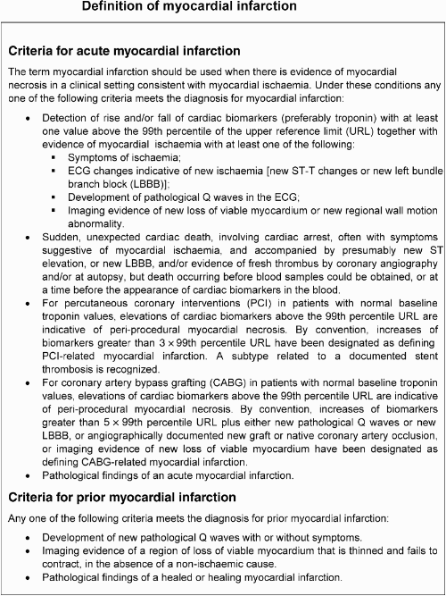
Clinically the various types of myocardial infarction can be classified as shown in Table 1.
Clinical classification of different types of myocardial infarction
| Type 1 Spontaneous myocardial infarction related to ischaemia due to a primary coronary event such as plaque erosion and/or rupture, fissuring, or dissection |
| Type 2 Myocardial infarction secondary to ischaemia due to either increased oxygen demand or decreased supply, e.g. coronary artery spasm, coronary embolism, anaemia, arrhythmias, hypertension, or hypotension |
| Type 3 Sudden unexpected cardiac death, including cardiac arrest, often with symptoms suggestive of myocardial ischaemia, accompanied by presumably new ST elevation, or new LBBB, or evidence of fresh thrombus in a coronary artery by angiography and/or at autopsy, but death occurring before blood samples could be obtained, or at a time before the appearance of cardiac biomarkers in the blood |
| Type 4a Myocardial infarction associated with PCI |
| Type 4b Myocardial infarction associated with stent thrombosis as documented by angiography or at autopsy |
| Type 5 Myocardial infarction associated with CABG |
On occasion, patients may manifest more than one type of myocardial infarction simultaneously or sequentially. It should also be noted that the term myocardial infarction does not include myocardial cell death associated with mechanical injury from coronary artery bypass grafting (CABG), for example ventricular venting, or manipulation of the heart; nor does it include myocardial necrosis due to miscellaneous causes, e.g. renal failure, heart failure, cardioversion, electrophysiological ablation, sepsis, myocarditis, cardiac toxins, or infiltrative diseases.
Biomarker evaluation
Myocardial cell death can be recognized by the appearance in the blood of different proteins released into the circulation from the damaged myocytes: myoglobin, cardiac troponin T and I, CK, LDH, as well as many others.3 Myocardial infarction is diagnosed when blood levels of sensitive and specific biomarkers such as cardiac troponin or CKMB are increased in the clinical setting of acute myocardial ischaemia.1 Although elevations in these biomarkers reflect myocardial necrosis, they do not indicate its mechanism.3,4 Thus, an elevated value of cardiac troponin in the absence of clinical evidence of ischaemia should prompt a search for other aetiologies of myocardial necrosis, such as myocarditis, aortic dissection, pulmonary embolism, congestive heart failure, renal failure, and other examples indicated in Table 2.
Elevations of troponin in the absence of overt ischemic heart disease
| Cardiac contusion, or other trauma including surgery, ablation, pacing, etc. |
| Congestive heart failure—acute and chronic |
| Aortic dissection |
| Aortic valve disease |
| Hypertrophic cardiomyopathy |
| Tachy- or bradyarrhythmias, or heart block |
| Apical ballooning syndrome |
| Rhabdomyolysis with cardiac injury |
| Pulmonary embolism, severe pulmonary hypertension |
| Renal failure |
| Acute neurological disease, including stroke or subarachnoid haemorrhage |
| Infiltrative diseases, e.g. amyloidosis, haemochromatosis, sarcoidosis, and scleroderma |
| Inflammatory diseases, e.g. myocarditis or myocardial extension of endo-/pericarditis |
| Drug toxicity or toxins |
| Critically ill patients, especially with respiratory failure or sepsis |
| Burns, especially if affecting >30% of body surface area |
| Extreme exertion |
The preferred biomarker for myocardial necrosis is cardiac troponin (I or T), which has nearly absolute myocardial tissue specificity as well as high clinical sensitivity, thereby reflecting even microscopic zones of myocardial necrosis.3 An increased value for cardiac troponin is defined as a measurement exceeding the 99th percentile of a normal reference population (URL = upper reference limit). Detection of a rise and/or fall of the measurements is essential to the diagnosis of acute myocardial infarction.6 The above-mentioned discriminatory percentile is designated as the decision level for the diagnosis of myocardial infarction, and must be determined for each specific assay with appropriate quality control.7–9 Optimal precision [coefficient of variation (CV)] at the 99th percentile URL for each assay should be defined as ≤10%. Better precision (CV ≤10%) allows for more sensitive assays.10,11 The use of assays that do not have independent validation of optimal precision (CV≤10%) is not recommended. The values for the 99th percentile can be found on the International Federation for Clinical Chemistry website http://www.ifcc.org/index.php?option=com_remository&Itemid=120&func=fileinfo&id=87.
Blood samples for the measurement of troponin should be drawn on first assessment (often some hours after the onset of symptoms) and 6–9 h later.12 An occasional patient may require an additional sample between 12 and 24 h if the earlier measurements were not elevated and the clinical suspicion of myocardial infarction is high.12 To establish the diagnosis of myocardial infarction, one elevated value above the decision level is required. The demonstration of a rising and/or falling pattern is needed to distinguish background elevated troponin levels, e.g. patients with chronic renal failure (Table 2), from elevations in the same patients which are indicative of myocardial infarction.6 However, this pattern is not absolutely required to make the diagnosis of myocardial infarction if the patient presents >24 h after the onset of symptoms. Troponin values may remain elevated for 7–14 days following the onset of infarction.4
If troponin assays are not available, the best alternative is CKMB (measured by mass assay). As with troponin, an increased CKMB value is defined as a measurement above the 99th percentile URL, which is designated as the decision level for the diagnosis of myocardial infarction.9 Gender-specific values should be employed.9 The CKMB measurements should be recorded at the time of the first assessment of the patient and 6–9 h later in order to demonstrate the rise and/or fall exceeding the 99th percentile URL for the diagnosis of myocardial infarction. An occasional patient may require an additional diagnostic sample between 12 and 24 h if the earlier CKMB measurements were not elevated and the clinical suspicion of myocardial infarction is high.
Measurement of total CK is not recommended for the diagnosis of myocardial infarction, because of the large skeletal muscle distribution and the lack of specificity of this enzyme.
Reinfarction
Traditionally, CKMB has been used to detect reinfarction. However, recent data suggest that troponin values provide similar information.13 In patients where recurrent myocardial infarction is suspected from clinical signs or symptoms following the initial infarction, an immediate measurement of the employed cardiac marker is recommended. A second sample should be obtained 3–6 h later. Recurrent infarction is diagnosed if there is a ≥20% increase of the value in the second sample. Analytical values are considered to be different if they are different by >3 SDs of the variance of the measures.14 For troponin, this value is 5–7% for most assays at the levels involved with reinfarction. Thus, a 20% change should be considered significant, i.e. over that expected from analytical variability itself. This value should also exceed the 99th percentile URL.
Electrocardiographic detection of myocardial infarction
The ECG is an integral part of the diagnostic work-up of patients with suspected myocardial infarction.1,2,15,16 The acute or evolving changes in the ST-T waveforms and the Q-waves when present potentially allow the clinician to date the event, to suggest the infarct-related artery, and to estimate the amount of myocardium at risk. Coronary artery dominance, size and distribution of arterial segments, collateral vessels, and location, extent, and severity of coronary stenoses can also impact ECG manifestations of myocardial ischaemia.17 The ECG by itself is often insufficient to diagnose acute myocardial ischaemia or infarction since ST deviation may be observed in other conditions such as acute pericarditis, LV hypertrophy, LBBB, Brugada syndrome, and early repolarization patterns.18 Also Q-waves may occur due to myocardial fibrosis in the absence of coronary artery disease, as in, for example, cardiomyopathy.
ECG abnormalities of myocardial ischaemia that may evolve to myocardial infarction
ECG abnormalities of myocardial ischaemia or infarction may be inscribed in the PR segment, the QRS complex, and the ST segment or T-waves. The earliest manifestations of myocardial ischaemia are typical T-waves and ST segment changes.19,20 Increased hyper-acute T-wave amplitude with prominent symmetrical T-waves in at least two contiguous leads is an early sign that may precede the elevation of the ST segment. Increased R-wave amplitude and width (giant R-wave with S-wave diminution) are often seen in leads exhibiting ST elevation, and tall T-waves reflecting conduction delay in the ischaemic myocardium.21 Transient Q-waves may be observed during an episode of acute ischaemia or rarely during acute myocardial infarction with successful reperfusion.22
Table 3 lists ECG criteria for the diagnosis of acute myocardial ischaemia that may lead to infarction. The J-point is used to determine the magnitude of the ST elevation. J-point elevation in men decreases with increasing age; however, that is not observed in women, in whom J-point elevation is less than in men.23
ECG manifestations of acute myocardial ischaemia (in absence of LVH and LBBB)
| ST elevation |
| New ST elevation at the J-point in two contiguous leads with the cut-off points: ≥0.2 mV in men or ≥0.15 mV in women in leads V2–V3 and/or ≥0.1 mV in other leads |
| ST depression and T-wave changes |
| New horizontal or down-sloping ST depression ≥0.05 mV in two contiguous leads; and/or T inversion ≥0.1 mV in two contiguous leads with prominent R-wave or R/S ratio >1 |
Contiguous leads means lead groups such as anterior leads (V1–V6), inferior leads (II, III, and aVF), or lateral/apical leads (I and aVL). More accurate spatial contiguity in the frontal plane can be established by the Cabrera display: aVL, I, aVR, II, aVF, and III.24 Supplemental leads such as V3R and V4R reflect the free wall of the right ventricle.
Although the criteria in Table 3 require that the ST shift be present in two or more contiguous leads, it should be noted that occasionally acute myocardial ischaemia may create sufficient ST segment shift to meet the criteria in one lead but have slightly less than the required ST shift in an adjacent contiguous lead. Lesser degrees of ST displacement or T-wave inversion in leads without prominent R-wave amplitude do not exclude acute myocardial ischaemia or evolving myocardial infarction.
ST elevation or diagnostic Q-waves in regional lead groups are more specific than ST depression in localizing the site of myocardial ischaemia or necrosis.25,26 However, ST depression in leads V1–V3 suggests myocardial ischaemia, especially when the terminal T-wave is positive (ST elevation equivalent), and may be confirmed by concomitant ST elevation ≥0.1 mV recorded in leads V7–V9.27,28 The term ‘posterior’ to reflect the basal part of the LV wall that lies on the diaphragm is no longer recommended. It is preferable to refer to this territory as inferobasal.29 In patients with inferior myocardial infarction it is advisable to record right precordial leads (V3R and V4R) seeking ST elevation in order to identify concomitant right ventricular infarction.30
During an acute episode of chest discomfort, pseudo-normalization of previously inverted T-waves may indicate acute myocardial ischaemia. Pulmonary embolism, intracranial processes, or peri-/myocarditis may also result in ST-T abnormalities and should be considered (false positives) in the differential diagnosis.
The diagnosis of myocardial infarction is difficult in the presence of LBBB even when marked ST-T abnormalities or ST elevation are present that exceed standard criteria.31,32 A previous ECG may be helpful to determine the presence of acute myocardial infarction in this setting. In patients with right bundle branch block (RBBB), ST-T abnormalities in leads V1–V3 are common, making it difficult to assess the presence of ischaemia in these leads; however, when ST elevation or Q-waves are found, myocardial ischaemia or infarction should be considered. Some patients present with ST elevation or new LBBB, and suffer sudden cardiac death before cardiac biomarkers become abnormal or pathological signs of myocardial necrosis become evident at autopsy. These patients should be classified as having had a fatal myocardial infarction.
Prior myocardial infarction
As shown in Table 4, Q-waves or QS complexes in the absence of QRS confounders are usually pathognomonic of a prior myocardial infarction.33–35 The specificity of the ECG diagnosis for myocardial infarction is greatest when Q-waves occur in several leads or lead groupings. ST deviations or T-waves alone are non-specific findings for myocardial necrosis. However, when these abnormalities occur in the same leads as the Q-waves, the likelihood of myocardial infarction is increased. For example, minor Q-waves ≥0.02 and <0.03 s that are ≥0.1 mV deep are suggestive of prior infarction if accompanied by inverted T-waves in the same lead group.
ECG changes associated with prior myocardial infarction
| Any Q-wave in leads V2–V3 ≥0.02 s or QS complex in leads V2 and V3 |
| Q-wave ≥0.03 s and ≥0.1 mV deep or QS complex in leads I, II, aVL, aVF, or V4–V6 in any two leads of a contiguous lead grouping (I, aVL,V6; V4–V6; II, III, and aVF)a |
| R-wave ≥0.04 s in V1–V2 and R/S ≥1 with a concordant positive T-wave in the absence of a conduction defect |
aThe same criteria are used for supplemental leads V7–V9, and for the Cabrera frontal plane lead grouping.
Other validated myocardial infarction-coding algorithms, such as the Minnesota code, Novacode, and WHO MONICA, define Q-wave depth on the basis of depth, width, and ratio of R-wave amplitude, such as Q-wave depth at least one-third or one-fifth of R-wave amplitude, and have been used extensively in epidemiological studies and clinical trials.36,37
Conditions that confound the ECG diagnosis of myocardial infarction
A QS complex in lead V1 is normal. A Q-wave <0.03 s and <1/4 of the R-wave amplitude in lead III is normal if the frontal QRS axis is between 30 and 0°. The Q-wave may also be normal in aVL if the frontal QRS axis is between 60 and 90°. Septal Q-waves are small non-pathological Q-waves <0.03 s and <1/4 of the R-wave amplitude in leads I, aVL, aVF, and V4–V6. Pre-excitation, obstructive or dilated cardiomyopathy, LBBB, RBBB, left anterior hemiblock, left and right ventricular hypertrophy, myocarditis, acute cor pulmonale, or hyperkalaemia may be associated with Q/QS complexes in the absence of myocardial infarction. ECG abnormalities that simulate myocardial ischaemia or infarction are presented in Table 5.
Common ECG pitfalls in diagnosing myocardial infarction
| False positives |
| Benign early repolarization |
| LBBB |
| Pre-excitation |
| Brugada syndrome |
| Peri-/myocarditis |
| Pulmonary embolism |
| Subarachnoid haemorrhage |
| Metabolic disturbances such as hyperkalaemia |
| Failure to recognize normal limits for J-point displacement |
| Lead transposition or use of modified Mason–Likar configuration24 |
| Cholecystitis |
| False negatives |
| Prior myocardial infarction with Q-waves and/or persistent ST elevation |
| Paced rhythm |
| LBBB |
Reinfarction
The ECG diagnosis of reinfarction following the initial infarction may be confounded by the initial evolutionary ECG changes. Reinfarction should be considered when ST elevation ≥0.1 mV reoccurs in a patient having a lesser degree of ST elevation or new pathognomonic Q waves, in at least two contiguous leads, particularly when associated with ischaemic symptoms for 20 min or longer. The re-elevation of the ST segment can, however, also be seen in threatening myocardial rupture and should lead to additional diagnostic work-up. ST depression or LBBB on their own should not be considered valid criteria for myocardial infarction.
Coronary revascularization
ECG abnormalities during or after percutaneous coronary intervention (PCI) are similar to those seen during spontaneous myocardial infarction. In patients who have undergone CABG, new ST-T abnormalities are common but not necessarily diagnostic of myocardial ischaemia.38 However, when new pathological Q waves (Table 4) appear in territories other than those identified before surgery, myocardial infarction should be considered, particularly if associated with elevated biomarkers, new wall motion abnormalities, or haemodynamic instability.
Imaging techniques
Non-invasive imaging plays many roles in patients with known or suspected myocardial infarction, but this section concerns only its role in the diagnosis and characterization of infarction. The underlying rationale is that regional myocardial hypoperfusion and ischaemia lead to a cascade of events including myocardial dysfunction, cell death, and healing by fibrosis. Important imaging parameters are therefore perfusion, myocyte viability, myocardial thickness, thickening, and motion, and the effects of fibrosis on the kinetics of radiolabelled and paramagnetic contrast agents.
Commonly used imaging techniques in acute and chronic infarction are echocardiography, radionuclide ventriculography, myocardial perfusion scintigraphy (MPS), and magnetic resonance imaging (MRI). Positron emission tomography (PET) and X-ray computed tomography (CT) are less common. There is considerable overlap in their capabilities, but only the radionuclide techniques provide a direct assessment of myocardial viability because of the properties of the tracers used. Other techniques provide indirect assessments of myocardial viability, such as myocardial function from echocardiography or myocardial fibrosis from MRI.
Echocardiography
Echocardiography is an excellent real-time imaging technique with moderate spatial and temporal resolution. Its strength is the assessment of myocardial thickness, thickening, and motion at rest. This can be aided by tissue Doppler imaging. Echocardiographic contrast agents can improve endocardial visualization, but contrast studies are not yet fully validated for the detection of myocardial necrosis, although early work is encouraging.39
Radionuclide imaging
Several radionuclide tracers allow viable myocytes to be imaged directly, including thallium-201, technetium-99m MIBI, tetrofosmin, and [18F]2-fluorodeoxyglucose (FDG).40–42 The strength of the techniques are that they are the only commonly available direct methods of assessing viability, although the relatively low resolution of the images disadvantages them for detecting small areas of infarction.43 The common single photon-emitting radio-pharmaceuticals are also tracers of myocardial perfusion and so the techniques readily detect areas of infarction and inducible perfusion abnormalities. ECG-gated imaging provides a reliable assessment of myocardial motion, thickening, and global function.44,45
Magnetic resonance imaging
Cardiovascular MRI has high spatial resolution and moderate temporal resolution. It is a well-validated standard for the assessment of myocardial function and has, in theory, similar capability to echocardiography in suspected acute infarction. It is, however, more cumbersome in an acute setting and is not commonly used. Paramagnetic contrast agents can be used to assess myocardial perfusion and the increase in extracellular space associated with the fibrosis of chronic infarction. The former is not yet fully validated in clinical practice, but the latter is well validated and can play an important role in the detection of infarction.46,47
X-Ray computed tomography
Infarcted myocardium is initially visible to CT as a focal area of decreased LV enhancement, but later imaging shows hyperenhancement as with late gadolinium imaging by MRI.48,49 This finding is clinically relevant because contrast-enhanced CT may be performed for suspected embolism and aortic dissection, conditions with clinical features that overlap with those of acute myocardial infarction.
Application in the acute phase of myocardial infarction
Imaging techniques can be useful in the diagnosis of myocardial infarction because of the ability to detect wall motion abnormalities in the presence of elevated cardiac biomarkers. If for some reason biomarkers have not been measured or may have normalized, demonstration of new loss of myocardial viability alone in the absence of non-ischaemic causes meets the criteria for myocardial infarction. However, if biomarkers have been measured at appropriate times and are normal, the determinations of these take precedence over the imaging criteria.
Echocardiography provides assessment of many non-ischaemic causes of acute chest pain such as peri-myocarditis, valvular heart disease, cardiomyopathy, pulmonary embolism, or aortic dissection. Echocardiography is the imaging technique of choice for detecting complications of acute infarction including myocardial free wall rupture, acute ventricular septal defect, and mitral regurgitation secondary to papillary muscle rupture or ischaemia. However, echocardiography cannot distinguish regional wall motion abnormalities due to myocardial ischaemia from infarction.
Radionuclide assessment of perfusion at the time of patient presentation can be performed with immediate tracer injection and imaging that can be delayed for up to several hours. The technique is interpreter dependent, although objective quantitative analysis is available. ECG gating provides simultaneous information on LV function.
An important role of acute echocardiography or radionuclide imaging is in patients with suspected myocardial infarction and a non-diagnostic ECG. A normal echocardiogram or resting ECG-gated scintigram has a 95–98% negative predictive value for excluding acute infarction.50–54 Thus, imaging techniques are useful for early triage and discharge of patients with suspected myocardial infarction.55,56
A regional myocardial wall motion abnormality or loss of normal thickening may be caused by acute myocardial infarction or by one or more of several other ischaemic conditions including old infarction, acute ischaemia, stunning, or hibernation. Non-ischaemic conditions such as cardiomyopathy and inflammatory or infiltrative diseases can also lead to regional loss of viable myocardium or functional abnormality, and so the positive predictive value of imaging techniques is not high unless these conditions can be excluded and unless a new abnormality is detected or can be presumed to have arisen in the setting of other features of acute myocardial infarction.
Application in the healing or healed phase of myocardial infarction
Imaging techniques are useful in myocardial infarction for analysis of LV function, both at rest and during dynamic exercise or pharmacological stress, to provide an assessment of remote inducible ischaemia. Echocardiography and radionuclide techniques, in conjunction with exercise or pharmacological stress, can identify ischaemia and myocardial viability. Non-invasive imaging techniques can diagnose healing or healed infarction by demonstrating regional wall motion, thinning, or scar in the absence of other causes.
The high resolution of contrast-enhanced MRI means that areas of late enhancement correlate well with areas of fibrosis and thereby enable differentiation between transmural and subendocardial scarring.57 The technique is therefore potentially valuable in assessing LV function and areas of viable and hence potentially hibernating myocardium.
Myocardial infarction associated with revascularization procedures
Peri-procedural myocardial infarction is different from spontaneous infarction, because the former is associated with the instrumentation of the heart that is required during mechanical revascularization procedures by either PCI or CABG. Multiple events that can lead to myocardial necrosis are taking place, often in combination, during both types of intervention.58–61 While some loss of myocardial tissue may be unavoidable during procedures, it is likely that limitation of such damage is beneficial to the patient and their prognosis.62
During PCI, myocardial necrosis may result from recognizable peri-procedural events, alone or in combination, such as side-branch occlusion, disruption of collateral flow, distal embolization, coronary dissection, slow flow or no-reflow phenomenon, and microvascular plugging. Embolization of intracoronary thrombus or atherosclerotic particulate debris cannot be entirely prevented despite current antithrombotic and antiplatelet adjunctive therapy or protection devices. Such events induce extensive inflammation of non-infarcted myocardium surrounding small islets of myocardium necrosis.63–67 New areas of myocardial necrosis have been demonstrated by MRI following PCI.68 A separate subcategory of myocardial infarction is related to stent thrombosis as documented by angiography and/or autopsy.
During CABG, numerous additional factors can lead to peri-procedural necrosis. These include direct myocardial trauma from sewing needles or manipulation of the heart, coronary dissection, global or regional ischaemia related to inadequate cardiac protection, microvascular events related to reperfusion, myocardial damage induced by oxygen free radical generation, or failure to reperfuse areas of the myocardium that are not subtended by graftable vessels.69–71 MRI studies suggest that most necrosis in this setting is not focal, but diffuse and localized to the subendocardium.72 Some clinicians and clinical investigators have preferred using CKMB for the diagnosis of peri-procedural infarction because of a substantial amount of data relating CKMB elevations to prognosis.73,74 However, an increasing number of studies using troponins in that respect have emerged.59,75
Diagnostic criteria for myocardial infarction with PCI
In the setting of PCI, the balloon inflation during a procedure almost always results in ischaemia whether or not accompanied by ST-T changes. The occurrence of procedure-related cell necrosis can be detected by measurement of cardiac biomarkers before or immediately after the procedure, and again at 6–12 and 18–24 h.76,77 Elevations of biomarkers above the 99th percentile URL after PCI, assuming a normal baseline troponin value, are indicative of post-procedural myocardial necrosis. There is currently no solid scientific basis for defining a biomarker threshold for the diagnosis of peri-procedural myocardial infarction. Pending further data, and by arbitrary convention, it is suggested to designate increases more than three times the 99th percentile URL as PCI-related myocardial infarction (type 4a).
If cardiac troponin is elevated before the procedure and not stable for at least two samples 6 h apart, there are insufficient data to recommend biomarker criteria for the diagnosis of peri-procedural myocardial infarction.77 If the values are stable or falling, criteria for reinfarction by further measurement of biomarkers together with the features of the ECG or imaging can be applied.
A separate subcategory of myocardial infarction (type 4b) is related to stent thrombosis as documented by angiography and/or autopsy. Although iatrogenic, myocardial infarction type 4b with verified stent thrombosis must meet the criteria for spontaneous myocardial infarction as well.
Diagnostic criteria for myocardial infarction with CABG
Any increase of cardiac biomarkers after CABG indicates myocyte necrosis, implying that an increasing magnitude of biomarker is likely to be related to an impaired outcome. This has been demonstrated in clinical studies employing CKMB where elevations five, 10 and 20 times the upper limit of normal after CABG were associated with worsened prognosis.73,78,79 Likewise, the increase of troponin levels after CABG indicates necrosis of myocardial cells, which predicts a poor outcome, in particular when elevated to the highest quartile or quintile of the troponin measurements.59,75
Unlike the prognosis, scant literature exists concerning the use of biomarkers for defining myocardial infarction in the setting of CABG. Therefore, biomarkers cannot stand alone in diagnosing myocardial infarction (type 5). In view of the adverse impact on survival observed in patients with significant biomarker elevations, this Task Force suggests, by arbitrary convention, that biomarker values more than five times the 99th percentile of the normal reference range during the first 72 h following CABG, when associated with the appearance of new pathological Q-waves or new LBBB, or angiographically documented new graft or native coronary artery occlusion, or imaging evidence of new loss of viable myocardium, should be considered as diagnostic of a CABG-related myocardial infarction (type 5 myocardial infarction).
Definition of myocardial infarction in clinical investigations
A universal definition for myocardial infarction would be of great benefit to future clinical studies in this area since it will allow for trial-to-trial comparisons as well as accurate meta-analyses involving multiple investigations. In clinical trials, myocardial infarction may be an entry criterion or an end-point. The definition of myocardial infarction employed in these trials will thus determine the characteristics of patients entering the studies as well as the number of outcome events. In recent investigations, different infarct definitions have been employed, thereby hampering comparison and generalization among these trials.
Consistency among investigators and regulatory authorities with regard to the definition of myocardial infarction used in clinical investigations is essential. The Task Force strongly encourages trialists to employ the definition described in this document. Furthermore, investigators should ensure that a trial provides comprehensive data for the various types of myocardial infarction (e.g. spontaneous, peri-procedural) and includes the employed decision limits for myocardial infarction of the cardiac biomarkers in question. All data should be made available to interested individuals in published format or on a website. Data concerning infarctions should be available in a form consistent with the current revised definitions of myocardial infarction. This does not necessarily restrict trialists to a narrow end-point definition, but rather ensures that across all future trials access to comparable data exists, thereby facilitating cross-study analyses. The recommendations put forward in this section are not detailed and should be supplemented by careful protocol planning and implementation in any clinical trial.
The Task Force strongly endorses the concept of the same decision limit for each biomarker employed for myocardial infarction types 1 and 2, and, likewise, the same higher three- and five-fold decision limits in the setting of myocardial infarction types 4a and 5, respectively78–80 (Tables 6 and 7). In clinical trials, as in clinical practice, measurement of cardiac troponin T or I is preferred over measurement of CKMB or other biomarkers for the diagnosis of myocardial infarction. Assessment of the quantity of myocardial damage (infarct size) is also an important trial end-point. Although the specific measurements vary depending on the assay and whether cardiac troponin T or I is used, in most studies troponin values correlate better with radionuclide- and MRI-determined infarct size than do CK and CKMB.81–83
Classification of the different types of myocardial infarction according to multiples of the 99th percentile URL of the applied cardiac biomarker
| Multiples × 99% . | MI type 1 (spontaneous) . | MI type 2 (secondary) . | MI type 3* (sudden death) . | MI type 4a** (PCI) . | MI type 4b (stent thrombosis) . | MI type 5** (CABG) . | Total number . |
|---|---|---|---|---|---|---|---|
| 1–2 × |  |  | |||||
| 2–3× | |||||||
| 3–5 × | |||||||
| 5–10 × | |||||||
| >10 × | |||||||
| Total number | |||||||
| Multiples × 99% . | MI type 1 (spontaneous) . | MI type 2 (secondary) . | MI type 3* (sudden death) . | MI type 4a** (PCI) . | MI type 4b (stent thrombosis) . | MI type 5** (CABG) . | Total number . |
|---|---|---|---|---|---|---|---|
| 1–2 × |  |  | |||||
| 2–3× | |||||||
| 3–5 × | |||||||
| 5–10 × | |||||||
| >10 × | |||||||
| Total number | |||||||
*Biomarkers are not available for this type of myocardial infarction since the patients expired before biomarker determination could be performed.
**For the sake of completeness, the total distribution of biomarker values should be reported. The dark grey areas represent biomarker elevations below the decision limit used for these types of myocardial infarction.
Irrespective of the specific end-point definition chosen in a clinical trial, all data should be provided. All boxes in the table should be completed, including the shaded areas.
Classification of the different types of myocardial infarction according to multiples of the 99th percentile URL of the applied cardiac biomarker
| Multiples × 99% . | MI type 1 (spontaneous) . | MI type 2 (secondary) . | MI type 3* (sudden death) . | MI type 4a** (PCI) . | MI type 4b (stent thrombosis) . | MI type 5** (CABG) . | Total number . |
|---|---|---|---|---|---|---|---|
| 1–2 × |  |  | |||||
| 2–3× | |||||||
| 3–5 × | |||||||
| 5–10 × | |||||||
| >10 × | |||||||
| Total number | |||||||
| Multiples × 99% . | MI type 1 (spontaneous) . | MI type 2 (secondary) . | MI type 3* (sudden death) . | MI type 4a** (PCI) . | MI type 4b (stent thrombosis) . | MI type 5** (CABG) . | Total number . |
|---|---|---|---|---|---|---|---|
| 1–2 × |  |  | |||||
| 2–3× | |||||||
| 3–5 × | |||||||
| 5–10 × | |||||||
| >10 × | |||||||
| Total number | |||||||
*Biomarkers are not available for this type of myocardial infarction since the patients expired before biomarker determination could be performed.
**For the sake of completeness, the total distribution of biomarker values should be reported. The dark grey areas represent biomarker elevations below the decision limit used for these types of myocardial infarction.
Irrespective of the specific end-point definition chosen in a clinical trial, all data should be provided. All boxes in the table should be completed, including the shaded areas.
Sample clinical trial tabulation of randomized patients by types of myocardial infarction
| Types of MI . | Treatment A . | Treatment B . |
|---|---|---|
| . | Number of patients . | Number of patients . |
| MI type 1 | ||
| MI type 2 | ||
| MI type 3 | ||
| MI type 4a | ||
| MI type 4b | ||
| MI type 5 | ||
| Total number |
| Types of MI . | Treatment A . | Treatment B . |
|---|---|---|
| . | Number of patients . | Number of patients . |
| MI type 1 | ||
| MI type 2 | ||
| MI type 3 | ||
| MI type 4a | ||
| MI type 4b | ||
| MI type 5 | ||
| Total number |
The use of cardiac troponins will undoubtedly increase the number of events recorded in a particular investigation because of increased sensitivity for detecting infarction.84–87 Ideally, data should be presented so that future clinical investigations or registries can translate the myocardial infarction end-point chosen in one study into the end-point of another study. Thus, measurements should be presented in a uniform manner to allow independent judgement and comparison of the clinical end-points. Furthermore, this Task Force suggests that data be reported as multiples of the 99th percentile URL of the applied biomarker, enabling comparisons between various classes and severity of the different types of myocardial infarction as indicated in Tables 6 and 7.
It is recommended that within a clinical trial all investigators whenever possible should employ the same assay in order to reduce the inter-assay variability, and, even better, the latter could be reduced to zero by application of a core laboratory using the same assay for all measurements.
In the design of a study, investigators should specify the expected effect of the new treatment under investigation. Factors that should be considered include:
Assessment of the incidence of spontaneous myocardial infarction (type 1) and infarction related to myocardial oxygen supplies and demand (type 2) in treated patients vs. control subjects.
Assessment of the incidence of sudden death related to myocardial infarction when applying the suggested criteria (type 3).
Assessment of the incidence of procedure-related myocardial infarctions and biomarker elevations (PCI, types 4a and 4b; and CABG, type 5).
Public policy implications of redefinition of myocardial infarction
Evolution of the definition of a specific diagnosis such as myocardial infarction has a number of implications for individual citizens as well as for society at large. The process of assigning a specific diagnosis to a patient should be associated with a specific value for the patient. The resources spent on recording and tracking a particular diagnosis must also have a specific value to society in order to justify the effort. A tentative or final diagnosis is the basis for advice about further diagnostic testing, treatment, lifestyle changes, and prognosis for the patient. The aggregate of patients with a particular diagnosis is the basis for health care planning and policy, and resource allocation.
One of the goals of good clinical practice is to reach a definitive and specific diagnosis, which is supported by current scientific knowledge. The approach to the definition of myocardial infarction outlined in this document meets this goal. In general, the conceptual meaning of the term myocardial infarction has not changed, although new sensitive diagnostic methods have been developed to diagnose this entity. Thus, the current diagnosis of acute myocardial infarction is a clinical diagnosis based on patient symptoms, ECG changes, and highly sensitive biochemical markers, as well as information gleaned from various imaging techniques. However, it is important to characterize the extent of the infarct as well as residual LV function and the severity of coronary artery disease, rather than merely making a diagnosis of myocardial infarction. The information conveyed about the patient's prognosis and ability to work requires more than just the mere statement that the patient has suffered an infarct. The multiple other factors just mentioned are also required so that appropriate social, family, and employment decisions can be made. A number of risk scores have been developed predicting post-infarction prognosis. The classification of the various other prognostic entities associated with myocardial necrosis should lead to a reconsideration of the clinical coding entities currently employed for patients with the myriad conditions that can lead to myocardial necrosis with consequent elevation of biomarkers.
Many patients with myocardial infarction die suddenly. Difficulties in definition of sudden and out-of-hospital death make attribution of the cause of death variable among physicians, regions, and countries. For example, out-of-hospital death is generally ascribed to ischaemic heart disease in the USA but to stroke in Japan. These arbitrary and cultural criteria need re-examination.
It is important that any revised criteria for the definition of myocardial infarction involve comparability of this definition over time so that adequate trend data can be obtained. Furthermore, it is essential to ensure widespread availability and standard application of the measures in order to ensure comparability of data from various geographic regions. Shift in criteria resulting in a substantial increase or decrease in case identification will have significant health resource and cost implications.86,87 Moreover, an increase in sensitivity of the criteria for myocardial infarction might entail negative consequences for some patients who are not currently labelled as having had an infarction. On the other hand, increasing diagnostic sensitivity for myocardial infarction can have a positive impact for a society: It should be appreciated that the agreed modification of the definition of myocardial infarction may be associated with consequences for the patients and their families with respect to psychological status, life insurance, professional career, as well as driving and pilot licences. Also the diagnosis is associated with societal implications as to diagnosis-related coding, hospital reimbursement, mortality statistics, sick leave, and disability attestation.
Increasing the sensitivity of diagnostic criteria for myocardial infarction will result in more cases identified in a society, thereby allowing appropriate secondary prevention.
Increasing the specificity of diagnostic criteria for myocardial infarction will result in more accurate diagnosis but will not exclude the presence of coronary artery disease, the cases of which may benefit from secondary prevention.
In order to meet this challenge, physicians must be adequately informed of the altered diagnostic criteria. Educational materials will need to be created and treatment guidelines must be appropriately adapted. Professional societies should take steps to facilitate the rapid dissemination of the revised definition to physicians, other health care professionals, administrators, and the general public.
Global perspectives of the redefinition of myocardial infarction
Cardiovascular disease is a global health problem. Approximately one-third of persons in the world die of cardiovascular disease, largely coronary artery disease and stroke, and 80% of these deaths from cardiovascular disease occur in developing countries. The greater proportion of deaths is due to heart disease and specifically coronary heart disease, of which myocardial infarction is a major manifestation. Since it is difficult to measure the prevalence of coronary artery disease in a population, the incidence of myocardial infarction may be used as a proxy, provided that a consistent definition is used when different populations, countries, or continents are being compared.
The changes in the definition of myocardial infarction have critical consequences for less developed and developing countries. In many countries, the resources to apply the new definition may not be available in all hospitals. However, many developing countries already do have medical facilities capable of or currently employing the proposed definition of myocardial infarction. In the context of the overall cost for a patient with myocardial infarction, the expense associated with a troponin assay would not be excessive and should be economically affordable in many hospitals in developing countries, particularly those where infarcts are frequent events. The necessary equipment, staff training, and running costs may be lacking in some regions, but certainly not in others. In less advantaged hospitals, the diagnosis of myocardial infarction may depend mostly on clinical signs and symptoms coupled with less sophisticated biomarker analyses. Some of these institutions may only have access to CK and its isoenzymes at the present time. The redefinition arises from and is compatible with the latest scientific knowledge and with advances in technology, particularly with regard to the use of biomarkers, high quality electrocardiography, and imaging techniques. The definition can and should be used by developed countries immediately, and by developing countries as quickly as resources become available.
The change in the definition of myocardial infarction will have a substantial impact on the identification, prevention, and treatment of cardiovascular disease throughout the world. The new definition will impact epidemiological data from developing countries relating to prevalence and incidence. The simultaneous and continuing use of the older WHO definition for some years would allow a comparison between data obtained in the past and data to be acquired in the future employing the newer biomarker approach. Cultural, financial, structural, and organizational problems in the different countries of the world in diagnosis and therapy of acute myocardial infarction will require on-going investigation. It is essential that the gap between therapeutic and diagnostic advances be addressed in this expanding area of cardiovascular disease.
Conflicts of interest
The members of the Task Force established by the ESC, the ACCF, AHA and the WHF have participated independently in the preparation of this document, drawing on their academic and clinical experience and applying an objective and critical examination of all available literature. Most have undertaken and are undertaking work in collaboration with industry and governmental or private health providers (research studies, teaching conferences, consultation), but all believe such activities have not influenced their judgement. The best guarantee of their independence is in the quality of their past and current scientific work. However, to ensure openness, their relations with industry, government and private health providers are reported in the ESC and European Heart Journal websites (www.escardio.org and www.eurheartj.org). Expenses for the Task Force/Writing Committee and preparation of this document were provided entirely by the above mentioned joint associations.
Acknowledgements
We are indebted to Professor Erling Falk MD for reviewing the section on ‘Pathology’. Furthermore, we are very grateful to the dedicated staff of the Guidelines Department of the ESC. We would also like to thank and to acknowledge the contribution of the following companies through unrestricted educational grants, none of whom were involved in the development of this publication and in no way influenced its contents. Premium observer: GSK. Observer: AstraZeneca, Beckman-Coulter, Dade Behring, Roche Diagnostics, Sanofi-Aventis, Servier.
References
The CME Text ‘Universal definition of myocardial infarction’ is accredited by the European Board for Accreditation in Cardiology (EBAC) for ‘1’ hour of external CME credits. Each participant should claim only those hours of credit that have actually been spent in the educational activity. EBAC works according to the quality standards of the European Accreditation Council for Continuing Medical Education (EACCME), which is an institution of the European Union of Medical Specialists (UEMS). In compliance with EBAC/EACCME guidelines, all authors participating in this programme have disclosed potential conflicts of interest that might cause a bias in the article. The Organizing Committee is responsible for ensuring that all potential conflicts of interest relevant to the programme are declared to the participants prior to the CME activities.
CME questions for this article are available at: European Heart Journalhttp://cme.oxfordjournals.org/cgi/hierarchy/oupcme_node;ehj and European Society of Cardiologyhttp://www.escardio.org/knowledge/guidelines.
Author notes
The recommendations set forth in this report are those of the Task Force Members and do not necessarily reflect the official position of the American College of Cardiology.
Dr Shanti Mendis of the WHO participated in the task force in her personal capacity, but this does not represent WHO approval of this document at the present time.
This article has been copublished in the October II (Vol. 28 no. 20), 2007, issue of the European Heart Journal (also available on the Web site of the European Society of Cardiology at www.escardio.org), the November 27, 2007, issue of Circulation (also available on the Web site of the American Heart Association at my.americanheart.org), and the November 27, 2007, issue of the Journal of the American College of Cardiology (also available on the Web site of the American College of Cardiology at www.acc.org).
This document was approved by the European Society of Cardiology in April 2007, the World Heart Federation in April 2007, and by the American Heart Association Science Advisory and Coordinating Committee May 9, 2007. The European Society of Cardiology, the American College of Cardiology, the American Heart Association, and the World Heart Federation request that this document be cited as follows: Thygesen K, Alpert JS, White HD; Joint ESC/ACCF/AHA/WHF Task Force for the Redefinition of Myocardial Infarction. Universal definition of myocardial infarction. Eur Heart J 2007;28:2525–2538.
The content of this European Society of Cardiology (ESC) document has been published for personal and educational use only. No commercial use is authorized. No part of the document may be translated or reproduced in any form without written permission from the ESC. Permission can be obtained upon submission of a written request to Oxford University Press, the publisher of the European Heart Journal and the party authorized to handle such permissions on behalf of the ESC.
Disclaimer. The document represents the views of the ESC, which were arrived at after careful consideration of the available evidence at the time they were written. Health professionals are encouraged to take them fully into account when exercising their clinical judgement. The document does not, however, override the individual responsibility of health professionals to make appropriate decisions in the circumstances of the individual patients, in consultation with that patient, and where appropriate and necessary the patient's guardian or carer. It is also the health professional's responsibility to verify the rules and regulations applicable to drugs and devices at the time of prescription.


