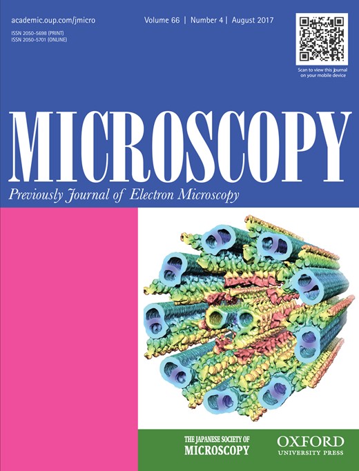
-
Views
-
Cite
Cite
Masao Yamashita, Matthew Ryan Leyden, Hidehito Adaniya, Martin Philip Cheung, Teruhisa Hirai, Yabing Qi, Tsumoru Shintake, Graphene specimen support technique for low voltage STEM imaging, Microscopy, Volume 66, Issue 4, August 2017, Pages 261–271, https://doi.org/10.1093/jmicro/dfx014
Close - Share Icon Share
Abstract
The scanning transmission electron microscopy (STEM) mode of today's field emission scanning electron microscopes enables sub-nanometer resolution imaging. Graphene is a single-atom thick, electrically conductive material, making it an excellent specimen support for the low voltage STEM imaging of nanometer-sized objects such as viruses. Here we present low voltage STEM images of bacteriophage T4 recorded on highly cleaned graphene films. The results show that ultrathin graphene support films markedly improve image signal at low accelerating voltages. Staining with a low atomic number methylamine vanadate stain combined with the graphene support film enables the clear visualization of the fine structure of the T4 tail by the low voltage STEM technique. Despite the advantages of graphene support films, difficulties are often encountered in placing hydrophilic biological samples on hydrophobic graphene electron microscopy grids. We employed a spin sedimentation sample loading method to overcome this problem.


