


Abstract
Primary sarcomas of the sellar region are uncommon, although a wide variety have been reported. To date, no cases of primary synovial sarcoma have been described as occurring at this site. We report an immunohistochemically and molecular genetically confirmed primary synovial sarcoma involving the sellar/parasellar region and cavernous sinus in an adult male. Subtotal resection and radiosurgery proved to be efficacious. The spectrum of primary sellar region sarcomas is summarized.
Skull base involvement by malignant tumors is usually the result of direct extension or metastatic disease. Well-recognized primary sarcomas involving this site, particularly the sella, include chordoma, chondrosarcoma, osteosarcoma, and a variety of postirradiation tumors, mainly fibrosarcomas. Pertinent to this report is but a single case of soft tissue synovial sarcoma metastatic to the sella.1 In this case report, we describe the first example of primary synovial sarcoma involving the sella/parasellar region, as well as the cavernous sinus. The morphologic diagnosis of synovial sarcoma was confirmed by immunohistochemistry and molecular genetics, which showed the diagnostic X;18 translocation. The patient, an adult male, initially responded favorably to subtotal resection and radiosurgery but has experienced a recurrence.
Case Study
A 48-year-old Caucasian male presented to the neurosurgical clinic with a 3-month history of right-sided headache and blurred vision. Neurologic and visual field examinations revealed mild right sixth nerve palsy, ptosis, and left hemianopsia. General laboratory values were normal, as was anterior and posterior pituitary function. A CT scan revealed a 3.5-cm irregular, contrast-enhancing mass involving the sella and right parasellar region. Punctate tumoral calcification was noted (Fig. 1). Further, MRI evaluation better defined the mass and showed involvement of the right cavernous sinus. Right optic nerve and chiasmal compression was also noted, as was narrowing (up to 60%) of the cavernous carotid artery throughout its course (Fig. 2A-C). Nonetheless, an MR angiogram showed normal blood flow. A right pterional craniotomy with subtotal (90%) resection of the mass was performed to decompress the optic nerve and obtain a diagnosis. The procedure was performed without complication.
A diagnosis of synovial sarcoma was made (see below). Because synovial sarcomas are often metastatic, an extensive radiographic workup was conducted; no other lesions were identified. Given the aggressive nature of the tumor, a more radical excision was undertaken after balloon test occlusion of the right carotid artery. Although no cross-filling occurred, monitoring over the 20-minute period of occlusion revealed no neurologic effect. A right orbitozygomatic osteotomy and Dolenc procedure were performed.2-4 Proximal control of the carotid artery was achieved through exposure of the petrous apex. The third, fourth, and fifth cranial nerves were grossly invaded by tumor and could not be salvaged. The adventitia of the cavernous carotid artery was also grossly infiltrated but was not resected because it had remained patent. Postoperative MRI scans showed only focal residual tumor on the carotid artery. Adjunctive, single-dose, image-guided radiosurgery (1,800 cGy to the 50% isodose line; maximum dose, 3,600 cGy) to the residual tumor and its bed (treatment volume, 6.3 cm3) was undertaken. With the exception of right ophthalmoplegia, the patient remained neurologically intact for 11 months, with no evidence of regrowth on MRI scan. Thereafter, a recurrence was noted just outside the original radiation field involving the skull base, and an exophytic retroclival extension caused compression upon the anterior pons. He continued to remain neurologically stable. Two weeks later, he underwent a subtotal tumor resection via a retrosigmoid approach to decompress the brainstem. The patient has since experienced progressive dysphagia.
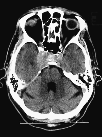
Contrast-enhanced Ct reveals a right sellar and parasellar mass. Calcification is seen in the lesion.
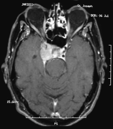
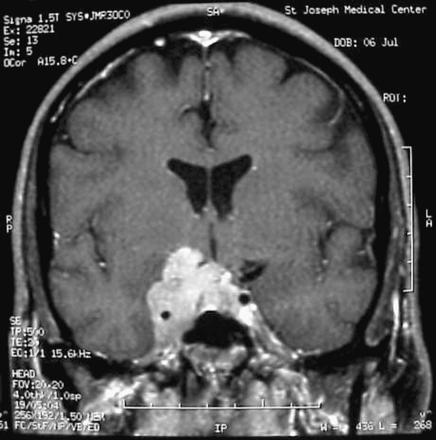
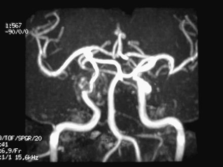
Axial contrast MRI shows a right sellar and parasellar mass (A). Coronal contrast MRI identifies optic nerve compression (B). MR angiography shows narrowing of the intracavernous carotid artery (C).
Materials and Methods
The formalin-fixed, routinely processed specimen was cut at 5 μm and stained with hematoxylin and eosin (H&E), Gomori reticulin, and periodic acid-Schiff with and without diastase digestion. Immunohistochemistry (streptavidin-biotin peroxidase complex method) utilized antisera directed against epithelial membrane antigen (EMA; Dako, Carpinteria, CA; 1:20, E29) keratin (AE1-AE3; Zymed, South San Francisco, CA; 1:200), vimentin (Dako; 1:500, 3B4), chromogranin (Roche, Indianapolis, IN; 1:1000, LK2H10), S-100 protein (Dako; 1:800, polyclonal), glial fibrillary acidic protein (Dako; 1:800, polyclonal), p53 protein (Dako; 1:200, DO-7), and Ki-67 (MIB-1; Dako; 1:800).
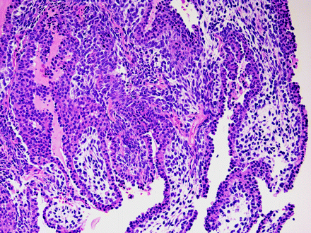
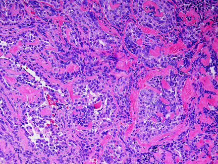
the histologically biphasic tumor featured a pseudopapillary epithelial component (A) and sheets of monotonous spindle cells enmeshed in collagen (B). Hematoxylin and eosin staining; original magnification ×160 (A) and ×250 (B).
RNA extraction and reverse transcriptase (RT)-PCR seeking synovial sarcoma fusion transcripts SYT-SSX1 and SYT-SSX2 were performed as previously described.5
Results
The first specimen consisted of fragments, the largest measuring 1.3 × 0.9 × 0.7 cm, and the second, 1 cm3 in aggregate. H&E-stained sections showed a biphasic, spindle- and epithelial-appearing tumor. The former consisted of cytologically uniform cells disposed in sheets and pseudopapillary arrangements in which an often single layer of neoplastic cells overlay a mucoid to fibrous, perivascular stoma (Fig. 3A). In other areas, sheets of tumor cells lay enmeshed in collagen (Fig. 3B). True glands were not seen. The spindle cell element was monomorphous. Few mitoses were noted (1/10 high-power fields at × × 40). No necrosis was encountered. Immunostains showed cytoplasmic staining for keratin (Fig. 4A) and membrane reactivity for EMA (Fig. 4B). All other markers but MIB-1 (labeling index, 22%) were immunonegative.
Molecular genetics studies (RT-PCR analysis) demonstrated the presence of the X;18 translocation (fusion transcript SYT-SSX2) diagnostic of synovial sarcoma.
Discussion
Synovial sarcoma is an uncommon soft tissue tumor, accounting for 5%-10% of malignant examples. Young adults are primarily affected, with a mean age of 30 years. Seventy-five percent occur near large joints, mainly of the lower extremities. Most of the remainder involve the trunk and the head and neck region with equal frequency,4 primarily the paravertebral and the retro- and parapharyngeal regions. Sinuses may also be affected.6
For the purpose of treatment planning, imaging of sarcomas of the cranial base requires both CT scan and MRI scanning. In the case of synovial sarcoma, a CT scan will identify calcifications in approximately one-third of cases.7 Only a minority are bone destructive. In soft tissue lesions, MRI shows variation in intensity and enhancement, as well as frequent septation.8 Most tumors are relatively iso- or hypointense compared to muscle and iso- or slightly hyperintense compared to fat and, in cystic areas, show a high fluid signal.9 If resection of the carotid artery is considered, angiography with balloon test occlusion should also be performed, which typically shows prominent vascularity.
Confirming the diagnosis of synovial sarcoma relies upon histologic and immunochemical features,10,11 and in some cases, molecular genetic analysis, as well.5 Three histologic patterns are recognized: the classic biphasic form with distinct epithelial and spindle cell components, and monophasic tumors of fibrous or epithelial type. The former occur with equal frequency, whereas the latter is rare. Our tumor is a biphasic example.
The growth rate of synovial sarcomas is often slow, belies their degree of malignancy, and precludes a timely diagnosis. The tumors are variably aggressive. Five-year survival rates cited often range from 40% to 75%.12-15 Local recurrence is frequent, and metastasis is common.14,16 Primary treatment of soft tissue examples involves aggressive resection with free margins.17-19 Unfortunately, skull base tumors may not be resectable. This is especially true of tumors of any kind involving the cavernous sinus region. Since synovial sarcoma rarely affects the sella, little is known regarding optimal treatment. One example secondarily affecting the parasellar area and orbit has been reported.1 It was a metastasis of a primary soft tissue monophasic synovial sarcoma of soft tissue operated on 16 months before. Treatment consisted of decompression of the cavernous sinus and radiation therapy. At 6-month follow-up, an MRI scan with contrast showed no tumor recurrence. In our case, an orbitozygomatic osteotomy combined with a Dolenc procedure allowed extensive bone removal and access to the cavernous sinus. Unfortunately, balloon test occlusion showed no cross-filling. Every attempt to preserve a tumor-free carotid artery was made, but due to adherence of residual tumor to its adventitia, a complete resection was not possible.
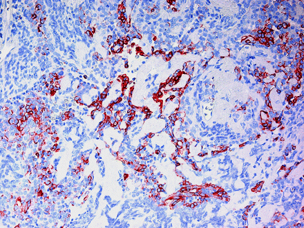
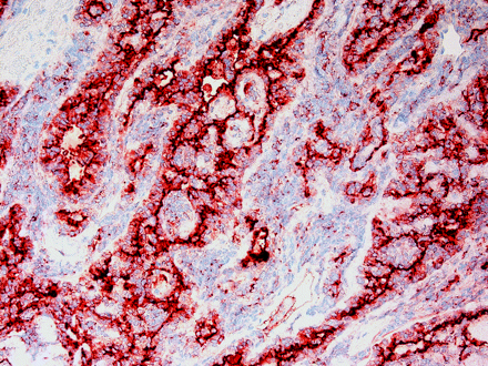
Immunohistochemistry revealed both cytoplasmic keratin reactivity (A) and membrane staining for epithelial membrane antigen (B). Original magnification ×250 for A and B.
Adjunctive treatments may be required. Radiation and chemotherapy have been studied and do result in local control.16,19-21 However, improved survival has not been seen.16,20,22 Advances in radiosurgery, specifically, image-guided robotic radiosurgery, allow for more accurate, aggressive treatment in the cranial base. No data are available to confirm whether this will be beneficial.
The lion's share of sellar tumors are pituitary adenomas. Their characteristic endocrine and radiographic abnormalities, coupled with lack of diabetes insipidus, distinguishes them for a wide variety of tumors affecting this anatomically complex region. Mesenchymal tumors occurring at this site include primarily meningiomas23 and hemangiopericytomas.24-27 The former are far more often benign or atypical than malignant,28 whereas the latter are malignant by definition, occurring in low- and high-grade forms.24,25,29 A variety of other benign and malignant mesenchymal tumors, arising either in dura or in bone, occasionally affect the sellar region.30,31 These include solitary fibrous tumor,32 fibrosarcoma,33-35 osteosarcoma,36 chondrosarcoma,37 mesenchymal chondrosarcoma,38 leiomyosarcoma,39,40 chordoma,41,42 rhabdomyosarcoma,43,44 and alveolar soft part sarcoma.45
A similar variety of sellar region sarcoma arises after radiotherapy, nearly always for pituitary adenoma. The relative and cumulative risk of their development has been calculated.46 The minimal therapeutic dose is approximately 3,000 cGy.47 The latency period from adenoma irradiation to presentation of the sarcoma varies greatly, ranging from as little as 2 years to as many as 40 years.48 Most examples are fibrosarcomas intimately associated with residual adenoma,47,49 but osteosarcoma48,50-52 and even leiomyosarcoma25 have also been reported. Fatal tumors often vary in their rate of progression and frequently recur but tend not to metastasize.
Conclusion
The spectrum of skull base sarcomas, particularly of the sellar region, is highly varied. Synovial sarcoma of the skull base is a rarely encountered entity. Most result in metastatic disease. This is the first reported case of synovial sarcoma involving the sella and parasellar region, as well as the cavernous sinus. The diagnosis was confirmed by both immunohistochemistry and molecular genetic testing. In that a complete resection could not be accomplished, it was felt that image-guided robotic radiosurgery would be appropriate, although its use has not previously been reported.
We appreciate the secretarial and photographic expertise of Denise Chase and James Hopfenspirger, respectively.
References
Buono LM, Silberschmidt A, Foroozan R, Savino PJ. Metastatic synovial sarcoma to the skull base and orbit.
Buster WP, Rodas RA, Fenstermaker RA, Kattner KA. Major venous sinus resection in the surgical treatment of recurrent aggressive dural based tumors.
Dolenc VV. Transcranial epidural approach to pituitary tumors extending beyond the sella.
Hakuba A, Liu S, Nishimura S. The orbitozygomatic infratemporal approach: a new surgical technique.
Jin L, Majerus J, Oliveira A, Inwards CY, Nascimento AG, Burgart LJ, Lloyd RV. Detection of fusion gene transcripts in fresh-frozen and formalin-fixed paraffin-embedded tissue sections of soft-tissue sarcomas after laser capture microdissection and Rt-PCR.
Gallia GL, Sciubba DM, Hann CL, et al. Synovial sarcoma of the frontal sinus. Case report.
McCarville MB, Spunt SL, Skapek SX, Pappo AS. Synovial sarcoma in pediatric patients.
van Rijswijk CS, Hogendoorn PC, Taminiau AH, Bloem JL. Synovial sarcoma: dynamic contrast-enhanced MR imaging features.
Jones BC, Sundaram M, Kransdorf MJ. Synovial sarcoma: MR imaging findings in 34 patients.
Ordonez NG, Mahfouz SM, Mackay B. Synovial sarcoma: an immunohistochemical and ultrastructural study.
Weiss SW, Goldblum TR. Malignant soft tissue tumors of uncertain types. In: Goldblum JR, Weiss SW, eds.
Brodsky JT, Burt ME, Hajdu SI, Casper ES, Brennan MF. Tendosynovial sarcoma. Clinicopathologic features, treatment, and prognosis.
Hajdu SI, Shiu MH, Fortner JG. Tendosynovial sarcoma: a clinicopathological study of 136 cases.
Lewis JJ, Antonescu CR, Leung DH, et al. Synovial sarcoma: a multivariate analysis of prognostic factors in 112 patients with primary localized tumors of the extremity.
Spillane AJ, A'Hern R, Judson IR, Fisher C, Thomas JM. Synovial sarcoma: a clinicopathologic, staging, and prognostic assessment.
Andrassy RJ, Okcu MF, Despa S, Raney RB. Synovial sarcoma in children: surgical lessons from a single institution and review of the literature.
Blakely ML, Spurbeck WW, Pappo AS, et al. The impact of margin of resection on outcome in pediatric nonrhabdomyosarcoma soft tissue sarcoma.
Raney RB. Synovial sarcoma in young people: background, prognostic factors, and therapeutic questions.
Pisters PW, Harrison LB, Leung DH, Woodruff JM, Casper ES, Brennan MF. Long-term results of a prospective randomized trial of adjuvant brachytherapy in soft tissue sarcoma.
Suit HD, Russell WO, Martin RG. Sarcoma of soft tissue: clinical and histopathologic parameters and response to treatment.
Okcu MF, Munsell M, Treuner J, et al. Synovial sarcoma of childhood and adolescence: a multicenter, multivariate analysis of outcome.
Solero CL, Giombini S, Morello G. Suprasellar and olfactory meningiomas. Report on a series of 153 personal cases.
Mena H, Ribas JL, Pezeshkpour GH, Cowan DN, Parisi JE. Hemangiopericytoma of the central nervous system: a review of 94 cases.
Niwa J, Hashi K, Minase t. Radiation induced intracranial leiomyosarcoma: its histopathological features.
Perry A, Scheithauer BW, Nascimento AG. the immunophenotypic spectrum of meningeal hemangiopericytoma: a comparison with fibrous meningioma and solitary fibrous tumor of meninges.
Perry A, Scheithauer BW, Stafford SL, Lohse CM, Wollan PC. “Malignancy” in meningiomas: a clinicopathologic study of 116 patients, with grading implications.
Perry A, Stafford SL, Scheithauer BW, Suman VJ, Lohse CM. Meningioma grading: an analysis of histologic parameters.
Ecker RD, Marsh WR, Pollock Be, et al. Hemangiopericytoma in the central nervous system: treatment, pathological features, and long-term follow up in 38 patients.
Radner H, Katenkamp D, Reifenberger G, Deckert M, Pietsch T, Wiestler OD. New developments in the pathology of skull base tumors.
Carneiro SS, Scheithauer BW, Nascimento AG, Hirose t, Davis DH. Solitary fibrous tumor of the meninges: a lesion distinct from fibrous meningioma. A clinicopathologic and immunohistochemical study.
Lopes MB, Lanzino G, Cloft HJ, Winston DC, Vance ML, Laws ER Jr. Primary fibrosarcoma of the sella unrelated to previous radiation therapy.
Moro M, Giannini C, Scheithauer BW, et al. Combined sellar fibrosarcoma and prolactinoma with neuronal metaplasia: report of a case unassociated with radiotherapy.
Nagasaka T, Nakashima N, Furui A, Wakabayashi T, Yoshida J. Sarcomatous transformation of pituitary adenoma after bromocriptine therapy.
Ashkan K, Pollock J, D'Arrigo C, Kitchen ND. Intracranial osteosarcomas: report of four cases and review of the literature.
Allan CA, Kaltsas G, Evanson J, et al. Pituitary chondrosarcoma: an unusual cause of a sellar mass presenting as a pituitary adenoma.
Inenaga C, Morii K, Tamura T, Tanaka R, Takahashi H. Mesenchymal chondrosarcoma of the sellar region.
Anderson WR, Cameron JD, Tsai SH. Primary intracranial leiomyosarcoma. Case report with ultrastructural study.
Kleinschmidt-DeMasters BK, Mierau GW, Sze CI, et al. Unusual dural and skull-based mesenchymal neoplasms: a report of four cases.
Mitchell A, Scheithauer BW, Unni KK, Forsyth PJ, Wold LE, McGivney DJ. Chordoma and chondroid neoplasms of the spheno-occiput. An immunohistochemical study of 41 cases with prognostic and nosologic implications.
Thodou E, Kontogeorgos G, Scheithauer BW, et al. Intrasellar chordomas mimicking pituitary adenoma.
Arita K, Sugiyama K, Tominaga A, Yamasaki F. Intrasellar rhabdomyosarcoma: case report.
Jalalah S, Kovacs K, Horvath E, Couldwell W, Weiss MH, Ezrin C. Rhabdomyosarcoma in the region of the sella turcica.
Bots GT, Tijssen CC, Wijnalda D, Teepen JL. Alveolar soft part sarcoma of the pituitary gland with secondary involvement of the right cerebral ventricle.
Minniti G, Traish D, Ashley S, Gonsalves A, Brada M. Risk of second brain tumor after conservative surgery and radiotherapy for pituitary adenoma: update after an additional 10 years.
Shi T, Farrell MA, Kaufmann JC. Fibrosarcoma complicating irradiated pituitary adenoma.
Florez JC, Burton DW, Arnell PM, Deftos LJ, Klibanski A. Hypercalcemia and local production of parathyroid hormone-related protein by a perisellar rhabdomyosarcoma after remote pituitary irradiation.
Prabhu SS, Aldape KD, Gagel RF, Benjamin RS, Trent JC, McCutcheon IE. Sarcomatous change after sellar irradiation in a growth hormone-secreting pituitary adenoma.
Bembo SA, Pasmantier R, Davis RP, Xiong Z, Weiss TE. Osteogenic sarcoma of the sella after radiation treatment of a pituitary adenoma.
Gnanalingham KK, Chakraborty A, Galloway M, Revesz T, Powell M. Osteosarcoma and fibrosarcoma caused by postoperative radiotherapy for a pituitary adenoma. Case report.


