


Abstract
The coronavirus disease 2019 (COVID-19) pandemic was declared a public health emergency of international concern by the World Health Organization. COVID-19 has high transmissibility and could result in acute lung injury in a fraction of patients. By counterbalancing the activity of the renin-angiotensin system, angiotensin-converting enzyme 2, which is the fusion receptor of the virus, plays a protective role against the development of complications of this viral infection. Vitamin D can induce the expression of angiotensin-converting enzyme 2 and regulate the immune system through different mechanisms. Epidemiologic studies of the relationship between vitamin D and various respiratory infections were reviewed and, here, the postulated mechanisms and clinical data supporting the protective role of vitamin D against COVID-19–mediated complications are discussed.
INTRODUCTION
Coronaviruses (CoVs) are positive, single-stranded RNA viruses capable of mutation and recombination and can infect mammals and birds, which can result in a spectrum of diseases.1–3 They are among the enveloped viruses with a small, membrane-integrated protein envelope that contributes to various aspects of the virus’s life cycle, including its assembly, budding, and pathogenesis,4 and makes them vulnerable to antimicrobial peptides.5 In humans, CoVs cause respiratory tract infections (RTIs) that are usually mild and are responsible for almost 15% of the epidemic common colds in winters. Coronaviruses also are able to contaminate a wide diversity of animals and cross species barriers, which results in outbreaks of severe (and sometimes lethal) RTIs such as severe acute respiratory syndrome (SARS) and the Middle East respiratory syndrome.1 In December 2019, Wuhan, China, became the origin of another outbreak of severe pneumonia (officially named coronavirus disease 2019 [COVID-19] by the World Health Organization) caused by a novel coronavirus called SARS-CoV-2.6–8 After an incubation period of 2–14 days (median, approximately 5 days),9 SARS-CoV-2 results in an RTI with manifestations ranging from asymptomatic or mild to a very severe form of the disease. The overall mortality rate among those hospitalized in China is 6.4%10 and the estimated mortality rate outside China is 15.2%11; the estimated basic reproduction number is between 2.24 and 3.58.2,12 As of this writing, most reported patients (mainly from China) are middle-aged adults and the disease seems to be more fatal in men and elderly patients.13–17 However, our knowledge regarding this disease is still in its infancy and rapidly evolving.
BACKGROUND
COVID-19 pathology
Applying a low-input, metagenomic, next-generation sequencing approach on RNA extracted from bronchoalveolar lavage of 2 patients with typical clinical features of the disease, Chen et al18 sequenced the entire genome of the SARS-CoV-2 virus encompassing 29 881 nucleotides.18 Advanced analyses demonstrated a 94.6% similarity between SARS-CoV-2 and SARS-CoV in the domains used for coronavirus species classification, suggesting the 2 viruses emerged from 1 species.19 More than a decade ago, it was demonstrated that SARS-CoV takes advantage of angiotensin-converting enzyme 2 (ACE2) as an essential receptor for binding to the host cells and infecting them.20 Importantly, Zhou et al,19 through a well-designed set of studies, demonstrated that SARS-CoV-2 also could use ACE2 as a receptor for cellular attachment in the ACE2-expressing cells of humans, civet, pig, and Chinese horseshoe bats (but not mouse), implying ACE2 is the fusion receptor of this virus.19
Apart from lung, ACE2 receptor is expressed in many other tissues, such as heart, endothelium, kidney, and intestine,21 which coincides with the tissue tropism of the SARS-CoV-2 virus. A review of preliminary studies showed that besides the main clinical findings (including fever, cough, and fatigue), acute respiratory distress, acute cardiac injury, acute renal failure, and diarrhea are among the most prevalent manifestations of COVID-19 infection.22,23 Angiotensin-converting enzyme 2 is the second angiotensin-converting enzyme and its main function is the regulation of the renin-angiotensin system (RAS).20 The RAS is a complex system consisting of a various enzymes, peptides, and receptors, including angiotensin-I-converting enzyme (ACE) and ACE2, and has an important role in several biological functions, such as regulation of the body blood pressure and water balance.24 The effects of RAS on lung injury could be mediated by its influence on the tone and permeability of lung vessels, vitality of epithelial cells, and the activation of fibroblasts.
Angiotensin-converting enzyme 2, in contrast to ACE, angiotensin (Ang) II, and Ang II type 1 receptor (AT1R), alleviated lung injury in experimental and in vivo models.25 Angiotensin-converting enzyme converts Ang I into Ang II, and binding of Ang II to AT1R contributes to a variety of consequences, such as vasoconstriction, proliferation, inflammation, and apoptosis. However, Ang-(1–7), a heptapeptide produced through hydrolysis of Ang II by ACE2, upon interaction with its receptor, neutralizes different functions of Ang II, conferring a counter-regulatory function for ACE2 against ACE.24,26 In 2002, in a candidate gene study, Marshall et al27 detected an association between the presence of a deletion polymorphism in the human ACE gene (DCP1), which is related to the higher activity of the ACE, with the development of acute respiratory distress syndrome and its associated mortality.27 After this study, Imai et al,28 in 2005, showed in an animal study that ACE2 can protect mice from development of sepsis-induced severe acute lung injury (ALI). In contrast, other constituents of the RAS (ie, ACE, Ang II, and AT1R) worsened the disease course and damaged pulmonary function. In this study, ACE-deficient mice demonstrated improvement in the disease manifestations, while a significant deterioration was detected in the lung performance of the Ace2 knockout mice. Also, the catalytic activity of ACE2 was suggested to protect against the development of ALI, whereas upregulation of Ang II by ACE was shown to play an important role in the pathogenesis of acute lung failure. On the other hand, the development of ALI was demonstrated to downregulate the ACE2 protein. Presumably, it was the first study that implied a direct protective role for ACE2 against the development of (sepsis-induced) lung injury.28 Later, these results were supported by other experimental studies that revealed a role for this enzyme in the protection against acute lung failure. In other words, ACE2 could be considered a key factor in both cell fusion of the virus and its pathogenicity, including lung, cardiovascular, and renal involvement.20
Vitamin D metabolism and deficiency
Although for years vitamin D was considered as a food constituent playing an important role in bone metabolism, to date, it is known as a steroid hormone with important regulatory roles in different physiological systems and pathways of the human body, including the immune system.29,30 Compelling evidence from around the world has illustrated an association between vitamin D deficiency and different infectious diseases, predominantly viral infections accompanied by a poor response to the standard treatments.30–34 Different clinical trials have demonstrated an association between vitamin D deficiency and increased risk of pulmonary infection, which is supported by various laboratory experimental studies highlighting different mechanisms, including the negative regulatory role of vitamin D on the renin-angiotensin signaling pathway. Vitamin D is produced in the skin from 7-dehydrocholesterol by ultraviolet radiation. Bound to the vitamin D–binding protein, vitamin D is transported to the liver, where it is hydroxylated to the major circulating form of vitamin D (25(OH)D) by at least 1 cytochrome P450 (CYP) hydroxylase. After transportation of 25(OH)D to the kidney, mediated by another CYP hydroxylase, the hormonally active form of vitamin D (1,25(OH)2D3) is synthesized.35,36 Although achieving the sufficient level of serum vitamin D is not far reaching, vitamin D deficiency is a global health issue. It is generally estimated that > 3 billion individuals around the world have vitamin D insufficiency, of whom half are suffering from actual vitamin D deficiency.37
ESTABLISHED CLINICAL EVIDENCE
Accumulating epidemiologic studies have demonstrated that low levels of plasma vitamin D could increase the incidence or severity of respiratory viral infections in humans, suggesting an important potential role for this vitamin in the prevention or treatment of viral RTIs.38 In a meta-analysis performed to study the association between low serum 25(OH)D concentration and RTIs in 11 randomized controlled trials (RCTs), including 5660 patients in total, Bergman et al39 reported a significant protective effect for vitamin D against RTI (odds ratio [OR], 0.64; 95%CI, 0.49–0.84). This protective effect was stronger when daily doses of vitamin D (vs bolus doses) were taken.39 In another meta-analysis, Pham et al40 discovered an inverse association between the circulating 25(OH)D level and risk (OR, 1.83; 95%CI, 1.42–2.37) as well as the severity (OR, 2.46; 95%CI, 1.65–3.66) of acute respiratory tract infections (ARTI). In this study, the highest risk of ARTI was associated with 25(OH)D concentrations < 15 ng/mL40 (to convert to nanomoles per liter, multiply by 2.496).
Vitamin D levels and dosing
Reviewing the current evidence suggests that the protective effect of vitamin D supplementation on reducing the incidence or severity of RTI only appears, or is most prominent in, patients deficient in vitamin D.41 Although, practically speaking, a serum 25(OH)D level < 20 ng/mL is considered deficient and < 30 ng/mL as insufficiency,42 in a secondary analysis of a US survey including 18 883 participants, 24% of individuals with 25(OH)D levels < 10 ng/mL reported having a recent RTI. In this study, cumulatively, recent RTI was reported by 44% of participants with vitamin D insufficiency, whereas only 17% of people with sufficient serum concentrations of vitamin D reported experiencing a recent RTI.43 Likewise, a case-control study of Bangladeshi children detected blood 25(OH)D concentrations of < 11.7 ng/mL to be linked with acute lower respiratory infection (ALRI) development. In this study, with each 4 ng/mL increase in the level of plasma vitamin D, the odds of ALRI development decreased by half.44 Also, in a case-control study conducted with 150 Indian children (aged 2–60 months), serum 25(OH)D concentrations > 9.01 ng/mL had a significantly protective effect against the development of severe ALRI (OR, 0.09; 95%CI, 0.03–0.24).45 In another Indian case-control study, in which the association between ALRI and serum vitamin D concentration was investigated, the mean ± SD serum 25(OH)D concentration of patients with ALRI (13.91 ± 2.99 ng/mL) was significantly less than that of the control subjects (19.38 ± 7.83 ng/mL).46 Also, in a Japanese case study of hospitalized children with ALRI, a significant correlation between serum vitamin D concentrations < 15 ng/mL and the need for ventilator management was detected.47 In addition, a hospital-based case-control study of Turkish newborns reported a significant difference between those with ALRI and healthy participants in terms of their own mean serum 25(OH)D level as well as their mothers’. In this study, neonates with blood 25(OH)D levels < 10 ng/mL (OR, 4.25; 95%CI, 1.05–17.07) compared with those with blood 25(OH)D concentrations < 20 ng/mL (OR, 1.83; 95%CI, 0.31–10.52) were more susceptible to ALRI development.48 The results of this study were confirmed by other studies of Turkish and Chinese infants.49,50 Moreover, in another neonatal study, the mean cord blood 25(OH)D level of neonates hospitalized because of an ALRI was significantly lower than that of the control subjects, who were newborns not hospitalized because of an ALRI (14.82 ng/mL vs 22.43 ng/mL).51
To date, the majority of the studies seeking an association between blood concentrations of vitamin D and ALI have been directed toward children. In 2 distinct meta-analyses, both published in 2017, by pooling the results of different studies of children, vitamin D deficiency was significantly associated with a higher risk for ALRI development.52,53 Furthermore, an association was detected between vitamin D deficiency and the severity of lower RTI in 1 of these studies.53 Finally, a meta-analysis on the results of 25 RCTs, including 10 933 individuals of all ages (range, 0 to 95 years), confirmed the association between vitamin D deficiency and ARTI. In this analysis, supplementation with vitamin D decreased the risk of ARTI among all study participants (OR, 0.88; 95%CI, 0.81–0.96). Through a subgroup analysis, Martineau et al54 confined the protective effect of vitamin D only for the daily or weekly intake of the supplement, in contrast to the bolus doses. Furthermore, within the group that took daily or weekly vitamin D supplementation, the best protective results were detected among individuals with 25(OH)D levels < 10.02 ng/mL. Martineau et al54 concluded that vitamin D was safe and protective against ARTI, with more prominent results in patients with severe vitamin D deficiency who received daily or weekly doses of the supplement. The results of this study were in line with the outcomes of a prior meta-analysis of RCTs published in 2013 by Bergman et al,39 who concluded that daily vitamin D supplementation was more protective compared with bolus doses (OR, 0.51 vs 0.86, respectively).39
Critically ill settings
Mortality rates have been reported to be higher in vitamin D–deficient patients in the medical intensive care unit (ICU) compared with patients with 25(OH)D insufficiency or those with sufficient 25(OH)D levels (mean ± SD 25(OH)D level of survivors vs nonsurvivors, 27.9 ± 9.7 vs 9.7 ± 4.7 ng/mL, respectively).55 Interestingly, another study conducted in a critical illness setting showed that although taking vitamin D during ICU hospitalization was not beneficial, patients who took vitamin D supplements before ICU admission experienced more ICU-free and ventilator-free days, indicating the importance of vitamin D supplementation in patients who may eventually need to be admitted to ICU.56 In an RCT conducted in 2016 in the United States, 30 patients in the ICU who were receiving mechanical ventilatory support were assigned to 1 of 3 groups: (1) placebo (n = 10), (2) 50 000 IU/d vitamin D3 for 5 sequential days (ie, total dose, 250 000 IU; n = 9), and (3) 100 000 IU/d vitamin D3 for 5 sequential days (ie, total dose, 500 000 IU; n = 11).57 In this study, administration of high doses of vitamin D3 safely increased the plasma concentration of 25(OH)D to the sufficient range (mean ± SD plasma 25(OH)D levels, 45.7 ± 19.6 ng/mL and 55.2 ± 14.4 ng/mL after the 250 000 IU and 500 000 IU vitamin D3 supplementations, respectively). Both regimens of vitamin D resulted in shortening hospitalization time. Although not significantly different, a decreasing trend was also noticed in length of ICU stay (mean ± SD, 23 ± 14 days for the placebo group vs 17 ± 14 and 15 ± 10 days in the 250 000 IU and 500 000 IU vitamin D3–supplementation groups, respectively) and in the number of days receiving ventilatory support (mean ± SD, 20 ± 15 in placebo vs 12 ± 10 and 14 ± 10 in the 250 000 IU and 500 000 IU vitamin D3–supplementation groups, respectively).57
Vitamin D supplementation and risk of toxicity
Ideally, the human body acquires a major part of its daily vitamin D need from certain foods, including dairy products, oily fish, and egg yolk, in addition to UVB-based synthesis in the skin.58,59 However, levels are not sufficient in a considerable part of the population worldwide and vitamin D deficiency is a global health issue for both sexes in all age groups.37,39 Furthermore, it should be noted that several groups of individuals, such as obese people, dark-skinned individuals living at high latitudes, seniors, patients with certain diseases such as renal disorders, as well as those who do not have enough vitamin D sources in their daily diet are at higher risk for vitamin D deficiency.55,60
To attain to an optimal 25(OH)D plasma level (30–50 ng/mL), international guidelines recommend daily supplementation of 400 IU–2000 IU for the general population (based on age, sex, body weight, skin color, time outdoors, and geographic latitude) throughout the year.58,59,61,62 Adults deficient in vitamin D are recommended to take weekly doses of 50 000 IU for up to 3 months or daily doses of 6000 IU followed by maintenance doses of 1500–2000 IU/d after achieving optimal blood concentration.58 The recommended doses increase for pregnant and breastfeeding women, obese adults (body mass index > 30 kg/m2), seniors, night workers, and dark-skinned people, as well as patients with a disability; and decrease for neonates and children.62,63 However, each country may have developed a clinical practice guideline with the recommendations best suited for its own residents.
Supplementation with both D2 (ergocalciferol) and D3 (cholecalciferol) is possible and the 2 types of the vitamin have been shown to be effective in maintaining the optimal level of vitamin D concentration.63,64 However, a study in 2004 of 20 healthy volunteers demonstrated a lower potency of less than one-third in addition to a shorter half-life for ergocalciferol relative to cholecalciferol.65 This result was confirmed by an RCT performed 15 years later.66 Therefore, it seems that D2 is less effective and has a shorter duration of action in humans compared with vitamin D3.65,66
A concern that may arise in this context is the possibility of vitamin D toxicity. Acute vitamin D toxicity, defined by blood 25(OH)D concentration > 150 ng/mL, is extremely rare and the acute toxic dose for vitamin D has not yet been well defined.67–69 On the basis of current evidence, the chronic toxicity appears after daily intake of > 40 000–60 000 IU for ≥ 1 months consecutively.67–71 Therefore, the possibility of vitamin D toxicity is slight. However, a report by the US Institute of Medicine in 2011, based on several association studies, suggested a possible association between serum 25(OH)D concentrations > 50 ng/mL and detrimental outcomes, such as increasing the risk of falls, certain cancers, and even death.69,72 In an observational cohort study with 5 years’ follow-up of approximately 250 000 Danish participants, the association between serum concentration of 25(OH)D and death due to cardiovascular disease, acute myocardial infarction, or stroke was examined. The lowest mortality risk was reported in association with vitamin D concentration of 22 ng/mL, whereas lower and higher extremes of vitamin D concentration were associated with increased mortality risk in a reverse J-shape (ie, the highest risk at the lower concentration levels). Compared with the optimal concentration (22 ng/mL), the vitamin D plasma level of 39.3 ng/mL was associated with a mortality hazard ratio of 1.3.73 However, in another observational study, analyzing data of 26 916 individuals from 8 European cohorts, increasing serum 25(OH)D concentrations up to 39.3 ng/mL was not associated with changes in the mortality rate.74 Nevertheless, 2 RCTs conducted with seniors ≥ 70 years demonstrated that high single doses of vitamin D (monthly or annually) could be related to a higher risk of falls and fractures in this age group.75,76 By and large, according to present knowledge, vitamin D toxicity could occur mainly as a result of taking extremely high doses of vitamin D and with the very high concentrations of blood 25(OH)D (ie, > 150 ng/mL).69
POTENTIAL MECHANISMS OF VITAMIN D IN MITIGATING COVID-19 PROGRESSION
RAS regulation
Various clinical and epidemiologic studies have demonstrated a strong link between vitamin D and the regulation of the RAS complex network.55,77–79 In lipopolysaccharide-induced ALI in rats, the active form of vitamin D can inhibit the biosynthesis of renin, ACE, and Ang II, and induce expression of ACE2. As a result, vitamin D, by inducing the activity of the ACE2/Ang-(1–7) axis and reducing renin and the activity of ACE/Ang II pathway, could negatively regulate the RAS.24 Blocking of the (pro)renin receptor in a randomized rodent model demonstrated protection against lipopolysaccharide-induced ALI by reduction of the inflammatory response in the pulmonary cells.80 A reduction in interstitial edema and hemorrhage, as well as a decline in the leukocyte count and levels of tumor necrosis factor-α and various interleukins, including interleukin-6, were also observed in this study.80 In addition, vitamin D could suppress the expression of renin, and this effect seems to be independent of the Ang II feedback regulation.77 In wild-type mice, vitamin D deficiency results in renin synthesis, and supplementation with 1,25(OH)2D3 suppresses the expression of renin.77 Furthermore, in an in vitro study using a cell line with high expression level of renin, Li et al77 found that 1,25(OH)2D3 directly and extensively suppressed the transcription of the renin gene by a vitamin D receptor (VDR)-mediated mechanism.77 Particularly, it has been observed that poor vitamin D status could result in increased susceptibility to infection by enveloped viruses.81 The nontrivial role played by the different elements of the RAS in the development of COVID-19 complications and the regulatory association between vitamin D and those elements underscore the importance of this postulated mechanism. However, in addition to the particular association between vitamin D and the CoV-specific receptor, ACE2, there are other interesting connections between this vitamin and the respiratory system and viral infections. Although the exact underlying mechanisms by which the poor vitamin D status could contribute to the development and worsening of the viral diseases remain to be fully discovered, there are a few hypotheses.82
Vitamin D receptor
Most of the immune system cells, including dendritic cells and T and B lymphocytes, express a high level of VDR, which functions as a transcription factor; upon binding to vitamin D, VDR modulates the responses of the cells to the viruses.61,81 Vitamin D receptor is also expressed in pulmonary tissue. In 2 distinct in vivo murine studies that compared the severity of lipopolysaccharide-induced ALI between VDR-knockout and wild-type mice, more severe ALI83,84 and higher mortality rate83 were reported in the VDR-knockout mice. Other important observations in the lung cells of VDR-knockout mice included elevated alveolar permeability and pulmonary vascular leakiness, increased neutrophil infiltration, apoptosis, pulmonary inflammation, and expression of pro-inflammatory cytokines and chemokines, in addition to the inordinate expression of Ang II.83,84 Moreover, FokI T allele was significantly associated with an increased viral infection susceptibility to the enveloped viruses.81
FokI is a major polymorphism in VDR that is associated with functional modifications in this receptor, such as inducing alterations in the interaction between VDR and its transcription factor that result in the changes in its transcriptional activity.81,85
Upregulation of the CYP27B1 gene and downstream antimicrobial peptides
In 2008, Hansdottir et al86 showed that primary lung epithelial cells express relatively high basal levels of CYP27B1 and low levels of CYP24A1. CYP27B1 encodes the 1α-hydroxylase enzyme that is responsible for the final step in the synthesis of the active, hormonal form of vitamin D (1,25(OH)2D3) from its circulating form (25(OH)D), whereas the CYP24A1 gene translates to 24-hydroxylase, which catalyzes the degradation of the active form of vitamin D. This locally generated, active vitamin D is believed to play an important role in the modulation of the immune system.86 Many immune cells, owing to the expression of CYP27B1, are also able to convert the inactive form of vitamin D to its active form. This activation is linked to the production and expression regulation of antimicrobial peptides such as α- and β-defensins and cathelicidins.30,87 This is one of the important roles played by vitamin D in the regulation of the immune system and confers critical antimicrobial functions and immunomodulatory activities in innate and adaptive immunity.88–93
Defensins are expressed by human airway epithelia and are present in airway secretions, playing a role in respiratory mucosal defence.94,95 Locally activated vitamin D can directly induce the expression of cathelicidin peptides.96 Intriguingly, Hansdottir et al,86 through a set of experiments on the live respiratory syncytial virus, detected that viral RNA is able to increase the expression of 1α-hydroxylase and, subsequently, conversion of vitamin D in primary epithelial cells, and they suggested the active form of vitamin D and viral RNA act synergistically to increase the expression of the antimicrobial peptides, particularly cathelicidin.86
Cathelicidins are antimicrobial peptides that are part of the innate immune system of many vertebrates, including humans, with a vast spectrum of direct and indirect antimicrobial activity against different pathogens, including enveloped viruses.97,98 The vitamin D-cathelicidin axis is considered an important player in the regulation of the human immune system and modulation of innate and adaptive immunity.99 LL-37 is the only identified member of the cathelicidin family in humans that is expressed by respiratory epithelial cells; it regulates and boosts the microbial combating capacity of the host cells against a wide range of respiratory pathogens.88,100,101 Vitamin D can induce the expression of the LL-37 gene (CAMP).102–104
Regulation of the inflammatory response
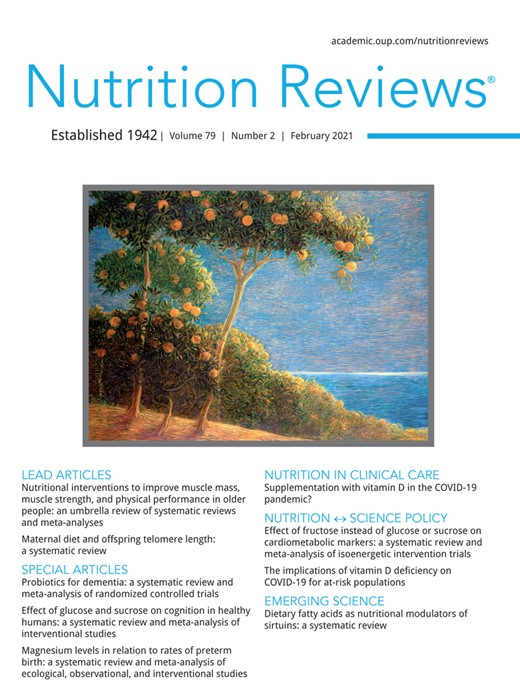
Regulating the activity of the important nuclear factor-κB (NF-κB) pathway, via various mechanisms, is among other suggested immunomodulatory functions of vitamin D.105–109 In an investigation of the influence of vitamin D on respiratory syncytial virus–infected airway epithelial cells in an in vitro model, researchers demonstrated that a locally produced active form of vitamin D could induce IκBα in the airway epithelium. IκBα is an inhibitor of NF-κB that can reduce the expression of the NF-κB–driven genes in response to the virus infection. IFN-b is one of these genes that translates to proteins with prominent antiviral activity. However, intriguingly, despite the reduction in the expression of IFN-b, this process does not increase the virus replication rate, implying it plays a role in controlling the inflammatory response of the body’s cells to the virus infection, which results in diminished severity of the disease and a lowering of the morbidity and mortality risks of the infection.38 Also, in a review article110 summarizing the results of in vitro investigations performed to explore the immunomodulatory effects of vitamin D in the virus-infected human respiratory epithelial cells, even though this vitamin and its metabolites were not shown to confer an inhibitory effect against virus replication in the cells, all the reviewed studies highlighted the influence of vitamin D on the expression and secretion of chemokines and pro-inflammatory cytokines. Although administration of vitamin D was associated with an increased secretion of the chemokines of CXCL8 and CXCL10 in rhinovirus-infected epithelial cells, vitamin D supplementation decreased the production of a wide range of different pro-inflammatory cytokines and chemokines in respiratory syncytial virus– and influenza A–infected cells.110 Postulated mechanisms through which vitamin D could play a protective role against viral RTIs are summarized in Figure 1.
![Postulated mechanisms through which vitamin D could play a protective role against viral RTI. (Upper box) Basal condition. Angiotensin (Ang) I, the product of the renin-mediated conversion of angiotensinogen, is hydrolyzed to Ang II by the peptidase angiotensin-I-converting enzyme (ACE). Ang II, upon binding to Ang II type 1 receptor (AT1R), confers its deleterious effects such as RTI through different mechanisms, including the development of interstitial edema and hemorrhage, and increasing the expression of pro-inflammatory cytokines and chemokines. Angiotensin-converting enzyme 2 (ACE2) hydrolyzes Ang II to Ang-(1–7), which upon interaction with its receptor (Mas), neutralizes different functions of Ang II and alleviates lung injury. IκBα is an inhibitor of NF-κB and reduces the expression of the NF-κB–driven genes (such as interferon β1 [IFN-b]) in response to the virus infection. IFN-b encodes a protein that, in response to a pathogen, induces transcription of inflammatory cytokines and chemokines. (Lower box) After treatment with vitamin D. Full arrowheads indicate the proposed effects of vitamin D. (A) The active form of vitamin D can suppress the expression of renin, ACE, and Ang II, and induce the expression of ACE2. (B) Suppression of the renin-angiotensin system could reduce the interstitial edema and hemorrhage and decrease the count of leukocytes and levels of pro-inflammatory cytokines (eg, TNF-α, IL-1β, IL-6, and IL-12) and pro-inflammatory transcription factors (eg, NF-κB). (C) The locally produced active form of vitamin D could induce IκBα in the airway epithelium which through inhibition of NF-κB reduces the expression of the NF-κB–driven genes in response to the virus infection. (D) Locally activated vitamin D can regulate the expression of certain host defense peptides (HDPs) such as cathelicidin, α- and β-defensins, and hepcidins. HDPs play a critical role in innate and adaptive immunity and through different mechanisms, such as reduction of the viral replication and infectivity,110 regulates and boosts the microbial combating capacity of the host against a wide range of the respiratory pathogens. *The CYP27B1 gene, which encodes 1α-hydroxylase, is highly expressed in many immune cells and primary lung epithelial cells. 1α-Hydroxylase catalyzes the final step in the synthesis of the active form of vitamin D3. Abbreviations: CYP27B1, cytochrome P450 family 27 subfamily b member 1; IL, interleukin; NF-κB, nuclear factor-κB; RTI, respiratory tract infection; TNF, tumor necrosis factor.](https://oup.silverchair-cdn.com/oup/backfile/Content_public/Journal/nutritionreviews/79/2/10.1093_nutrit_nuaa081/1/m_nuaa081f1.jpeg?Expires=1716358884&Signature=m~H9CsPGs0eC68p1SwKloYB4Vs8Eaot7ryvxPHlpr7ViB-vDrruAg8V9JHeL-64z0gPNGKzjvVnjGKYZAiUQ4i99ECBm-PvuC39d99lfC6f-sHitNBojIPusbWwF6BfHjYpDupIaNp84bD4vqGgxSmG28VK9admCTCE2NfGByAVKEZdQe5xf4eMpWSlyotz8-E9DjEF~kaYWANobVAgTPIwkkE115pE0vfq15N5fBfR5oxOEdEVlkZVI~AUimFvfuuDnaWilp~BJbv--FmOFDq0y6W0kG5hqSRKSHeHnXXSrMmx2J-vOfDvO4MsI6CedGP1mPQtNdR3yM6IC1yp96g__&Key-Pair-Id=APKAIE5G5CRDK6RD3PGA)
Postulated mechanisms through which vitamin D could play a protective role against viral RTI. (Upper box) Basal condition. Angiotensin (Ang) I, the product of the renin-mediated conversion of angiotensinogen, is hydrolyzed to Ang II by the peptidase angiotensin-I-converting enzyme (ACE). Ang II, upon binding to Ang II type 1 receptor (AT1R), confers its deleterious effects such as RTI through different mechanisms, including the development of interstitial edema and hemorrhage, and increasing the expression of pro-inflammatory cytokines and chemokines. Angiotensin-converting enzyme 2 (ACE2) hydrolyzes Ang II to Ang-(1–7), which upon interaction with its receptor (Mas), neutralizes different functions of Ang II and alleviates lung injury. IκBα is an inhibitor of NF-κB and reduces the expression of the NF-κB–driven genes (such as interferon β1 [IFN-b]) in response to the virus infection. IFN-b encodes a protein that, in response to a pathogen, induces transcription of inflammatory cytokines and chemokines. (Lower box) After treatment with vitamin D. Full arrowheads indicate the proposed effects of vitamin D. (A) The active form of vitamin D can suppress the expression of renin, ACE, and Ang II, and induce the expression of ACE2. (B) Suppression of the renin-angiotensin system could reduce the interstitial edema and hemorrhage and decrease the count of leukocytes and levels of pro-inflammatory cytokines (eg, TNF-α, IL-1β, IL-6, and IL-12) and pro-inflammatory transcription factors (eg, NF-κB). (C) The locally produced active form of vitamin D could induce IκBα in the airway epithelium which through inhibition of NF-κB reduces the expression of the NF-κB–driven genes in response to the virus infection. (D) Locally activated vitamin D can regulate the expression of certain host defense peptides (HDPs) such as cathelicidin, α- and β-defensins, and hepcidins. HDPs play a critical role in innate and adaptive immunity and through different mechanisms, such as reduction of the viral replication and infectivity,110 regulates and boosts the microbial combating capacity of the host against a wide range of the respiratory pathogens. *The CYP27B1 gene, which encodes 1α-hydroxylase, is highly expressed in many immune cells and primary lung epithelial cells. 1α-Hydroxylase catalyzes the final step in the synthesis of the active form of vitamin D3. Abbreviations: CYP27B1, cytochrome P450 family 27 subfamily b member 1; IL, interleukin; NF-κB, nuclear factor-κB; RTI, respiratory tract infection; TNF, tumor necrosis factor.
CONCLUSION
ACE2 is considered as receptor for SARS-CoV-2 through which the virus attaches to the cell. However, ACE2 also plays a protective role in the development of complicated manifestations of this viral infection.20 The active form of vitamin D can induce the expression of ACE224 and regulate the immune system through different distinct mechanisms. There is evidence from laboratory experiments that vitamin D can reduce the replication of other respiratory viruses and this is supported by clinical data demonstrating a preventive role for vitamin D against viral RTIs, particularly in those suffering from very low plasma concentrations of vitamin D. Taking this information together and regarding the important role of ACE2 in the prevention of COVID-19 complications on one hand and its regulatory association with vitamin D on the other hand, it seems rational to consider a potential role for vitamin D against SARS-Cov-2, although to date, we do not know for sure whether such an effect exists. Presumably, the most protective effect of supplementation appears in patients deficient in vitamin D (ie, those with serum 25(OH)D levels < 10 ng/mL) and possibly prior to the emergence of complications. Although vitamin D is safe (toxicity being quite rare), easily available, and affordable, its deficiency is a worldwide health problem.37 Therefore, supplementation with daily (preferred) or weekly doses of vitamin D, aimed at increasing the concentration of serum 25(OH)D to the optimal level of 30–50 ng/mL69,111–114 could be considered a global strategy, being more important in countries with a high prevalence of vitamin D deficiency and particularly in those patients with COVID-19 who are a high risk of ICU admission.
Acknowledgments
The author wishes to thank Prof. Joseph Rafter for kindly reviewing this manuscript.
Author contributions. F.H. is solely responsible for all aspects of this report and is accountable for all aspects of the work.
Funding. None.
Declaration of interest. None.
References
Du Y, Tu L, Zhu P, et al. Clinical Features of 85 Fatal Cases of COVID-19 from Wuhan. A Retrospective Observational Study. Am J Respir Crit Care Med. 2020;
