


Abstract
To provide an overview of a multisociety effort to formulate appropriate use criteria for image-guided injections and radiofrequency procedures in the diagnosis and treatment of sacroiliac joint and posterior sacroiliac complex pain.
The Spine Intervention Society convened a multisociety effort to guide physicians and define for payers the appropriate use of image-guided injections and radiofrequency procedures. An evidence panel was established to write systematic reviews, define key terms and assumptions, and develop clinical scenarios to be addressed. The rating panel considered the evidence presented in the systematic reviews, carefully reviewed the definitions and assumptions, and rated the clinical scenarios. Final median ratings, in combination with the level of agreement, determined the final ratings for the appropriate use of sacroiliac injections and radiofrequency neurotomy.
More than 10,000 scenarios were addressed in the appropriate use criteria and are housed within five modules in the portal, available on the Spine Intervention Society website: Module 1: Clinical Indications and Imaging; Module 2: Anticoagulants; Module 3: Timing of Injections; Module 4: Number of Injections; and Module 5: Lateral Branch Radiofrequency Neurotomy. Within several of these modules, several issues of interest are identified and discussed.
Physicians and payers can access the appropriate use criteria portal on the Spine Intervention Society’s website and select specific clinical indications for a particular patient in order to learn more about the appropriateness of the intervention(s) under consideration.
Introduction
Being an innervated structure [1–5], the sacroiliac joint is a potential source of pain. Noxious stimulation of the joint in normal volunteers evokes back pain [6–9], and clinical studies have shown the sacroiliac joint to be the source of pain in about one in five patients with chronic low back pain [10–12].
Likewise, the posterior ligaments of the sacroiliac joint are innervated [13] and are, therefore, a potential source of pain. Noxious stimulation of these ligaments evokes pain in normal volunteers [8,9], but no clinical studies have yet determined how often the posterior sacroiliac ligaments are the source of pain in patients with low back pain. Significantly for clinical purposes, studies have shown that local anesthetic blocks of the lateral branches of the sacral dorsal rami protect asymptomatic volunteers from noxious stimulation of the interosseous and dorsal sacroiliac ligaments, but not the sacroiliac joints [9].
Multiple studies have reported various success rates for relieving pain with injections of corticosteroids into the sacroiliac joint, but typically these studies had only a short duration of follow-up [12]. Success rates may have been overestimated in observational studies because such studies do not exclude the possibility of benefit from nonspecific or placebo effects [14]. On the other hand, in studies in which a valid diagnosis of sacroiliac joint pain was not previously made, success rates may have been underestimated by the inclusion of patients who do not have sacroiliac joint pain.
Several studies have attempted to relieve sacroiliac pain by performing radiofrequency neurotomy of the lateral branches of the sacral dorsal rami, with or without inclusion of the L5 dorsal ramus. For achieving at least 50% relief of pain, the reported success rate of this type of treatment is approximately 50% [15]. The majority of studies, however, selected subjects on the basis of their responses to intra-articular sacroiliac joint injections, rather than diagnostic blocks of the sacral lateral branches, which are the target of this therapeutic procedure; ironically, lateral branch blocks do not protect normal volunteers from sacroiliac joint pain.
Given these limitations in the literature, physicians are seeking guidance on how best to diagnose and treat SIJ and posterior sacroiliac complex pain, while insurers are wrestling with coverage decisions. For such situations, appropriate use criteria (AUC) can be developed in order to define areas of appropriate use, along with identifying potential overuse and underuse of procedures.
Methods
The objectives of the present AUC are 1) to provide physicians with a tool to assist in diagnosing and treating SIJ and posterior sacroiliac ligament pain utilizing image-guided injections and radiofrequency procedures and 2) to define for payers what is typically appropriate use of image-guided injections and radiofrequency procedures for these patients. This AUC does not address the entire spectrum of treatment options for sacroiliac pain.
The Appropriate Use Criteria Committee of the Spine Intervention Society adapted the RAND/UCLA Appropriateness Method (RAM) to guide development of appropriate use criteria [16]. RAM has been utilized extensively as a means to integrate the best available scientific evidence with the clinical judgment of experts.
Once the sacroiliac interventions topic was chosen, the Society invited other medical specialty societies, representing physicians involved in the care of patients with SIJ and posterior sacroiliac complex pain, to participate in a multisociety, multidisciplinary collaboration. The medical specialty societies that participated in the project with the Spine Intervention Society were the American Academy of Orthopaedic Surgeons, American Society of Anesthesiologists, American College of Radiology, American Academy of Physical Medicine and Rehabilitation, American Academy of Pain Medicine, and North American Spine Society. All invited societies appointed members to serve on both the evidence and rating panels.
The evidence panel was charged with 1) writing systematic reviews that summarized and evaluated the existing evidence [12,15]; 2) developing clinical scenarios that encompassed important clinical indications and interventional treatments to be evaluated by the rating panel (Appendix 1); and 3) formulating definitions (Appendix 2) and assumptions (Supplementary Data File S1, available online) to clarify terminology and scope. The rating panel was responsible for rating the clinical scenarios after carefully reviewing the definitions and assumptions and the evidence presented in the systematic reviews. All members of both panels disclosed potential conflicts of interest (Supplementary Data File S2, available online).
Two systematic reviews were completed in 2014 and served as the evidence base for the AUC project: One addressed diagnostic and therapeutic intra-articular sacroiliac injections [12], and the other addressed diagnostic and therapeutic posterior sacroiliac interventions, specifically lateral branch blocks and lateral branch radiofrequency neurotomy [15]. The authors of the two systematic reviews [12,15] appraised the evidence according to the Grading of Recommendations Assessment, Development and Evaluation (GRADE) system of evaluating evidence, and in both cases the body of evidence was found not to be of high quality.
Without a solid, high-quality evidence base, the rating panel members were reliant to a large extent upon their own clinical experience in assessing the clinical scenarios regarding the appropriateness of the diagnostic and therapeutic image-guided injections and radiofrequency procedures for patients presenting with various combinations of clinical indications. Given the number of clinical indications and interventions, the rating panel members independently assessed more than 10,000 clinical scenarios, twice.
Each scenario was rated on a scale of 1–9, on which a score of 1–3 indicates that the intervention is inappropriate for the given clinical indications; 4–6 denotes uncertainty; and 7–9 assesses the intervention as appropriate. Members of the rating panel rated the clinical scenarios once in March–April 2014, prior to a face-to-face meeting. Two weeks before the face-to-face meeting, members were provided with a report of their own ratings for each clinical scenario, along with anonymous ratings of the scenarios from the other members of the panel. The report also identified median ratings and whether there was agreement among reviewers.
The intention of the face-to-face meeting in May 2014 was to encourage discussion of scenarios with discrepant ratings or significant disagreement, not for the purpose of achieving consensus but in order to ensure that all members similarly understood the scenarios. Additionally, several definitions and many clinical scenarios were revised during the course of the meeting in order to reflect more accurately the intended indications referred to in the scenarios.
Following the meeting, members once again rated the scenarios in May–June 2014. The results of the second round of ratings were then circulated to the rating panel members for review and confirmation that their final, second round ratings accurately reflected their assessments, especially for the revised scenarios, which they had rated only once. The final median rating, in combination with the level of agreement, determined the final ratings for the appropriate use of sacroiliac injections and radiofrequency neurotomy.
Consistent with RAM, the definitions of levels of appropriateness and levels of agreement are as follows:
Levels of Appropriateness
Appropriate = panel median of 7–9, without disagreement
Uncertain = panel median of 4–6 OR any median with disagreement
Inappropriate = panel median of 1–3, without disagreement
Levels of Agreement (for Panels of 11–13 Members)
Agreement = no more than three panelists rate the appropriateness of the intervention for the scenario outside the three-point region (1–3, 4–6, 7–9) containing the median
Neutral = more than three panelists rate outside the three-point region, but fewer than four ratings in an alternate three-point region
Disagreement = four or more ratings in each extreme three-point region
Results
More than 10,000 scenarios were addressed in the AUC. It is not practical to present them all here. It is important, however, to provide an introduction to the five modules housed in the AUC Portal (Module 1: Clinical Indications and Imaging; Module 2: Anticoagulants; Module 3: Timing of Injections; Module 4: Number of Injections; Module 5: Lateral Branch Radiofrequency Neurotomy) and provide a breakdown of the indications and interventions contained in each module of the AUC (see Appendix 2). Within several of these modules, there are issues that merit some discussion and explanation.
Module 1: Clinical Indications and Imaging (Initial Injection)
The modules that address the appropriateness of sacroiliac injections and radiofrequency procedures for specific clinical indications and imaging are organized by primary location of pain, including pain localized to the SIJ, pain over the SIJ and referred into the leg, pain over the SIJ with referral into the groin, maximal ipsilateral pain above the L5 vertebra, and suspected acute spondyloarthritis. Within each module, important variables to consider comprise imaging findings, diagnostic physical examination testing, prior diagnostic injections, and potentially pertinent patient history.
When reviewing the location of pain as an independent variable, maximal pain above the L5 vertebra was negatively correlated with the recommendation for an SIJ injection. Other historical items, including the presence of spondyloarthritis, had minimal impact on the ratings. The rating panel placed more emphasis on physical examination findings. In scenarios with three or more positive provocation SIJ tests, the injection was given a high level of appropriateness regardless of the remainder of the scenario details. SIJ injections were also seen as appropriate for pain in the presence of one or two positive provocation tests depending on the other scenario variables. SIJ injections were not felt to be appropriate in subjects without a clinical exam or in those with no positive provocation maneuvers.
The rating panel placed little emphasis on imaging findings. There did not seem to be a clear distinction made between “degenerative changes” and “abnormal findings” on imaging studies despite these having been defined in the assumptions document. In fact, in some instances, when all other variables were equal, the presence of “degenerative” SIJ changes on imaging was more likely to generate a recommendation for an SIJ injection than the presence of “abnormal findings.” This is felt to be an inconsistency and is likely the result of rater fatigue or a misinterpretation of the definitions of these different imaging findings.
When considering an initial injection in this module, the rating panel preferred injections with a combination of local anesthetic and steroid to injections of local anesthetic alone. This is likely reflective of practice patterns within the United States, given that the majority of societies involved comprise practitioners from the United States; initial injections are discussed in more detail below (see Timing and Number of Injections). For the initial injections that were addressed in this module, there were no recommendations to inject steroid without local anesthetic. In addition, there were no clinical criteria for which the panel agreed that it was appropriate to perform lateral branch blocks as a first intervention.
Module 2: Anticoagulants
The rating panel made clear recommendations to not withhold anticoagulant or antiplatelet medications prior to injecting the SIJ or lateral branches. This is likely based on the lack of bleeding complications reported in the literature combined with the absence of sensitive neural structures that could be damaged by a hematoma if bleeding were to occur. When anticoagulant medication is withheld, there is likely to be a greater risk posed by the condition for which anticoagulants were prescribed.
Modules 3 and 4: Timing and Number of Injections
The rating panel concluded that intra-articular injections of local anesthetic and steroid are an appropriate first intervention when pain has been present for more than one month, has an intensity of greater than 4/10, and is causing functional limitations, regardless of whether or not conservative therapy had been provided. In general, injections were considered appropriate for pain of lesser intensity and duration if the pain was causing functional limitation and conservative treatment had been provided.
As in Module 1, there were no scenarios for which an intra-articular injection of steroid alone was considered an appropriate first intervention. Also similar to Module 1, the rating panel preferred the injection of local anesthetic and steroid to an injection of local anesthetic alone as an initial injection. The median rating for an initial injection of local anesthetic alone was, in general, 1 point lower than the injection of local anesthetic and steroid. This did result in some scenarios in which injections of local anesthetic and steroid were considered appropriate, but injections of local anesthetic alone were considered uncertain, or injections of local anesthetic and steroid were considered appropriate with agreement, whereas injections of local anesthetic alone were considered appropriate without agreement.
Based upon rating panel discussion, we hypothesize that the justification for this phenomenon lies not in any lesser degree of appropriateness of first proceeding with a diagnostic injection without steroid; rather, it likely reflects the desire to limit the number of injections administered to a single patient. Physicians who perform a first injection that includes steroid are aware that they are administering a therapeutic agent to a patient who has not yet been diagnosed with sacroiliac joint pain. If the response to local anesthetic is positive, then they have saved the patient a subsequent office visit for an additional therapeutic injection, thereby reducing the travel burden to the patient, exposure to radiation, and reducing the albeit small risk of an infection from a subsequent injection. However, if the patient has a negative response to the local anesthetic, they have been unnecessarily exposed to steroid. The apparent inconsistency may well be an unintended consequence of payer limitations on the number of injections that will be reimbursed for a given patient’s episode of care for suspected sacroiliac joint pain.
It was the opinion of the rating panel that injections of steroid with local anesthetic, injections of steroid alone, and lateral branch blocks would all be appropriate following an initial diagnostic injection that provided greater than 75% relief. Injections of local anesthetic and steroid were generally rated as more appropriate than other injections if the relief was greater than 50%. Further injections were generally not recommended if the pain relief was less than 50%.
The rating panel concluded that an injection of local anesthetic and steroid would be appropriate if there was at least 50% relief from an initial therapeutic injection or at least 75% relief from a subsequent injection, regardless of the duration of relief, and that an injection of steroid alone would only be appropriate if there was at least 75% relief for two months.
Module 5: Lateral Branch Radiofrequency Neurotomy
Two key factors were identified for the evaluation of indications for a lateral branch radiofrequency neurotomy (LBRFN): duration of symptoms and degree of pain relief obtained during blocks. The rating panel specified that patients should have symptoms for a minimum duration of two to three months prior to undergoing this procedure. Raters also clearly felt that obtaining less than 50% pain relief from diagnostic injections was insufficient justification to proceed with LBRFN. Increased percentage of pain relief and duration of symptoms both correlated with higher levels of appropriateness, although raters did not differentiate between 75% and 100% pain relief, which were treated as equivalent.
Similar trends emerged for consideration of repeat LBRFN. Repeat LBRFN was not deemed appropriate if the first LBRFN resulted in less than 50% pain relief or if the duration of effect was less than three months. Increasing the duration and percentage of pain relief resulted in higher levels of appropriateness, although the raters again did not discriminate between 75% and 100% pain relief. The type and sequence of block obtained (intra-articular vs lateral branch block) had minimal effect on the outcome and were most relevant for those with 50–75% pain relief and in those with only two to three months of symptoms.
Conclusion
Final ratings for the clinical scenarios are now available via a link to the AUC Portal of the Spine Intervention Society at http://www.spineintervention.org/?page=S1_AUC. Physicians can access the portal, review the assumptions and disclaimer, and proceed to select the module(s) of interest. By selecting the clinical indications for a particular patient, the physician will obtain information on the appropriateness of the intervention(s) under consideration. For those interested in reviewing the report that lists the median ratings and agreement for every clinical scenario, a PDF is available at http://www.spineintervention.org/?page=S1_AUC.
Acknowledgments
On behalf of the Spine Intervention Society, the authors would like to extend our deepest gratitude to all members of the evidence panel for their assistance in critically assessing the evidence, which served as the basis for the systematic reviews.
Evidence Panel Members: Drs. Anil Sharma, Chair (Spine Intervention Society), Shihab Ahmed (Spine Intervention Society), Thiru Annaswamy (American Academy of Physical Medicine and Rehabilitation), Jamie Baisden (North American Spine Society), Asokumar Buvanendran (American Society of Anesthesiologists), Michael DePalma (Spine Intervention Society), Andrew Engel (Spine Intervention Society), Wellington Hsu (American Academy of Orthopaedic Surgeons), Wade King (Spine Intervention Society), Tim Lamer (American Academy of Pain Medicine), Devi Nampiaparampil (Spine Intervention Society), Nileshkumar Patel (Spine Intervention Society), Jeffrey Peterson (American College of Radiology), Jeffrey Summers (Spine Intervention Society).
A special thanks also to members of the rating panel who spent countless hours considering the evidence and completing the ratings. Your stamina and patience have been greatly appreciated.
Rating Panel Members: Drs. Ray Baker (Spine Intervention Society), Asokumar Buvanendran (American Society of Anesthesiologists), Srinivas Chiravuri (Spine Intervention Society), Eduardo Fraifeld (American Academy of Pain Medicine), Mary Jesse (American College of Radiology), Milton Landers (Spine Intervention Society), Heidi Prather (North American Spine Society), Gwendolyn Sowa (American Academy of Physical Medicine and Rehabilitation), Claire Tibiletti (Spine Intervention Society), William C. Watters III (American Academy of Orthopaedic Surgeons).
Finally, the authors wish to thank Ms. Sandra Ray, Manager of Policy and Practice at the Society, for her assistance with managing the project.
Supplementary Data
Supplementary Data may be found online at http://painmedicine.oxfordjournals.org.
Appendix 1 Definition and Derivation of Clinical Scenarios
For each module, multiple individual hypothetical scenarios were created by systematically combining the clinical feature specified in the title of the module with each of the features listed under “indications” in the table for each module. In turn, each of the features in the first column of indications was combined with each of the features listed in any subsequent column. The number of scenarios thus developed for each module was the arithmetic product of the number of features listed in each column. For each scenario, assessors would rate the appropriateness of each of the procedures listed in the table.
1. Clinical Indications and Imaging
The patient has pain localized to the region of the sacroiliac joint
| Indications | Procedures | ||
|---|---|---|---|
| Imaging | Diagnostic Tests | History | |
|
|
|
|
| Indications | Procedures | ||
|---|---|---|---|
| Imaging | Diagnostic Tests | History | |
|
|
|
|
SIJ = sacroiliac joint.
The patient has pain localized to the region of the sacroiliac joint
| Indications | Procedures | ||
|---|---|---|---|
| Imaging | Diagnostic Tests | History | |
|
|
|
|
| Indications | Procedures | ||
|---|---|---|---|
| Imaging | Diagnostic Tests | History | |
|
|
|
|
SIJ = sacroiliac joint.
The patient has pain located over the sacroiliac joint and referred into the lower limb
| Indications | Procedures | ||
|---|---|---|---|
| Imaging | Diagnostic Tests | History | |
|
|
|
|
| Indications | Procedures | ||
|---|---|---|---|
| Imaging | Diagnostic Tests | History | |
|
|
|
|
SIJ = sacroiliac joint.
The patient has pain over the sacroiliac joint and in the groin
| Indications | Procedures | ||
|---|---|---|---|
| Imaging | Diagnostic Tests | History | |
|
|
|
|
| Indications | Procedures | ||
|---|---|---|---|
| Imaging | Diagnostic Tests | History | |
|
|
|
|
SIJ = sacroiliac joint.
The patient has pain over the sacroiliac joint and in the groin
| Indications | Procedures | ||
|---|---|---|---|
| Imaging | Diagnostic Tests | History | |
|
|
|
|
| Indications | Procedures | ||
|---|---|---|---|
| Imaging | Diagnostic Tests | History | |
|
|
|
|
SIJ = sacroiliac joint.
The patient has maximal ipsilateral pain above the level of the L5 vertebra
| Indications | Procedures | ||
|---|---|---|---|
| Imaging | Diagnostic Tests | History | |
|
|
|
|
| Indications | Procedures | ||
|---|---|---|---|
| Imaging | Diagnostic Tests | History | |
|
|
|
|
SIJ = sacroiliac joint.
The patient is suspected to have acute spondyloarthritis
| Indications | Procedures |
|---|---|
|
|
| Indications | Procedures |
|---|---|
|
|
SIJ = sacroiliac joint.
The patient is suspected to have acute spondyloarthritis
| Indications | Procedures |
|---|---|
|
|
| Indications | Procedures |
|---|---|
|
|
SIJ = sacroiliac joint.
2. Anticoagulation
The patient is taking anticoagulants
| Indications | Procedures |
|---|---|
|
|
| Indications | Procedures |
|---|---|
|
|
NSAID = nonsteroidal anti-inflammatory drug; SIJ = sacroiliac joint.
The patient is taking anticoagulants
| Indications | Procedures |
|---|---|
|
|
| Indications | Procedures |
|---|---|
|
|
NSAID = nonsteroidal anti-inflammatory drug; SIJ = sacroiliac joint.
3. Timing
The patient is being considered for an interventional procedure
| Indications | Procedures | ||
|---|---|---|---|
| Pain Severity | Duration | Conservative Treatment | |
|
|
|
|
| Indications | Procedures | ||
|---|---|---|---|
| Pain Severity | Duration | Conservative Treatment | |
|
|
|
|
SIJ = sacroiliac joint.
4. Number of Injections
The patient is being considered for a second intervention. A first injection produced relief of pain for the expected duration of action of the local anesthetic used
| Indications | Procedures |
|---|---|
| Degree of Relief | |
| <50% | Intra-articular SIJ injection of local anesthetic with steroid? |
| ≥50% | Intra-articular SIJ injection of local anesthetic without steroid? |
| ≥75% | Intra-articular SIJ injection of steroid alone? |
| 100% | Lateral branch blocks? |
SIJ = sacroiliac joint.
The patient is potentially eligible for an interventional procedure following dual diagnostic injections; each injection has provided relief of pain for the expected duration of action of the local anesthetic used
| Indications | Procedures | |||
|---|---|---|---|---|
| First Diagnostic Injection | Second Diagnostic Injection | |||
| Agents Used | Relief | Agents Used | Relief | |
| Local anesthetic |
| Local anesthetic |
|
|
| Local anesthetic with steroid |
| Local anesthetic with steroid |
| |
| Local anesthetic |
| Local anesthetic | None | |
| Local anesthetic with steroid |
| Local anesthetic with steroid | None | |
SIJ = sacroiliac joint.
The patient has had relief from a previous therapeutic injection and is being considered for a repeat therapeutic injection
| Indications | Procedures | ||
|---|---|---|---|
| Previous Injection | Relief | Duration of Relief | |
|
|
|
|
| Indications | Procedures | ||
|---|---|---|---|
| Previous Injection | Relief | Duration of Relief | |
|
|
|
|
SIJ = sacroiliac joint.
5. Lateral Branch Radiofrequency Neurotomy
The patient is being considered for lateral branch radiofrequency neurotomy. If performed, diagnostic blocks have provided relief for the expected duration of action of the local anesthetic used
| Indications | Procedure | ||||
|---|---|---|---|---|---|
| First Diagnostic Block | Second Diagnostic Block | Duration of Symptoms | |||
| Site | Relief | Site | Relief | ||
|
|
| Lateral branch radiofrequency neurotomy? | ||
|
| ||||
| Indications | Procedure | ||||
|---|---|---|---|---|---|
| First Diagnostic Block | Second Diagnostic Block | Duration of Symptoms | |||
| Site | Relief | Site | Relief | ||
|
|
| Lateral branch radiofrequency neurotomy? | ||
|
| ||||
SIJ = sacroiliac joint.
The patient has had relief from a previous lateral branch radiofrequency neurotomy and is being considered for repeat treatment
| Indications | Procedure | |
|---|---|---|
| Previous Relief | Duration of Relief | |
| <50% | <3 months | Lateral branch radiofrequency neurotomy? |
| ≥50% | 3–6 months | |
| ≥75% | 6–12 months | |
| 100% | >12 months | |
| Indications | Procedure | |
|---|---|---|
| Previous Relief | Duration of Relief | |
| <50% | <3 months | Lateral branch radiofrequency neurotomy? |
| ≥50% | 3–6 months | |
| ≥75% | 6–12 months | |
| 100% | >12 months | |
Appendix 2 Fluoroscopically Guided Diagnostic and Therapeutic Sacroiliac Interventions: Clinical Scenario Definitions
Anticoagulant medication: medications designed to prevent blood coagulation. These medications include coumarins (warfarin, acenocoumarol, phenprocoumon), heparin and derivatives (heparin, low–molecular weight heparins, fondaparinux, idraparinux), direct factor Xa inhibitors (rivaroxaban, apixaban), and direct thrombin inhibitors (e.g., dabigatran, hirudin, lepirudin, argatroban, dabigatran).
Antiplatelet agents: any medication designed to reduce platelet aggregation and inhibit thrombus formation. These medications include irreversible cyclooxygenase inhibitors (aspirin), adenosine diphosphate receptor inhibitors (ticlopidine, clopidogrel, prasugrel, etc.), phosphodiesterase inhibitors (cilostazol), glycoprotein IIB/IIIA inhibitors (e.g., abciximab, eptifibatide), adenosine reuptake inhibitors (dipyridamole), and thromboxane inhibitors.
Conservative treatment: for the purpose of this document, conservative treatment refers to medical treatment (e.g., nonsteroidal anti-inflammatory drugs, activity modification, physical therapy) designed to avoid more invasive interventional procedures.
Diagnostic spine injection(s): fluoroscopically guided interventional procedure(s) performed for the purpose of diagnosing the source of pain. In the lumbar spine, these include intra-articular zygapophysial joint injections, lumbar medial branch blocks, lumbar spinal nerve blocks, and provocation discography.
Diagnostic hip injection(s): injections of local anesthetic directed toward or into structures that are suspected to be sources of hip girdle pain (e.g., hip joint injection for intra-articular hip pathology, iliopsoas or trochanteric bursa injection for suspected bursitis).
Fluoroscopic guidance: use of fluoroscopy to guide the placement of needles and/or electrodes for invasive diagnostic and therapeutic procedures.
Fusion through L5-S1: any surgical procedure that involves fixating at least the lowest motion segment of the spine. This would include any discectomy procedure with interbody fusion, with or without the presence of posterior hardware (e.g., interspinous fixator, pedicle screws). In the case of anatomic variations (sacralized L5), fusion through L4-S1 would be included.
Hip pathology: any hip condition that can produce groin pain. This would include, but is not limited to, osteoarthritis of the hip, labral injuries, and iliopsoas bursitis.
Imaging: for the purposes of this document, imaging refers to any imaging modality that can adequately demonstrate pathology of the affected area. Examples would include plain radiographs, computed tomography scans, nuclear imaging (bone scan, SPECT), magnetic resonance imaging (typically with STIR images).
Recent imaging is defined as imaging obtained during the current episode to obtain information about the pathology of the affected area.
Degenerative changes on imaging are findings that may be related to an aging spine or joint that may or may not be symptomatic, including osteophytes, joint osteoarthrosis (or arthritis), disc desiccation and/or bulging, and loss of disc height. Findings on imaging that suggest pathological change may also be asymptomatic.
Abnormal findings on imaging of the lumbar spine might include acute fractures, acute disc protrusions or extrusions, high-intensity zones, bony edema presence on STIR or T2 fat saturated images, and/or positive bone scan with or without SPECT. In the case of patients with a prior L5-S1 fusion, abnormal imaging of the lumbar spine might include a pseudoarthrosis or adjacent-level disease.
Abnormal findings on pelvic imaging (includes bony pelvis, sacroiliac joint and related structures; excludes the hip joint) include bony edema presence on STIR or T2 fat saturated images and/or positive bone scan with or without SPECT.
Abnormal findings on imaging of the hip (includes acetabulum, hip joint, femoral head, and related structures) include radiographic findings consistent with full-thickness articular cartilage loss (subchondral cysts), severe osteoarthritis, labral injuries, iliopsoas bursitis, the presence of bony edema on STIR or T2 fat saturated images, and/or positive bone scan with or without SPECT.
Inciting event: traumatic or cumulative circumstance thought to be the cause of an injury.
Laboratory data: in the context of spondyloarthropathy, erythrocyte sedimentation rate and C-reactive protein levels are typically (though not always) elevated; a positive HLA-B27 is typical (though not diagnostic).
Lateral branch blocks (LBB): image-guided nerve blocks of the lateral sacral branches at S1–3, usually supplemented by an L5 dorsal ramus block.
Lateral branch radiofrequency neurotomy (LBRFN): image-guided thermal (not nonthermal or pulsed) ablation of the lateral sacral branches at S1–3, usually supplemented by ablation of the L5 dorsal ramus. For the purposes of this document, only radiofrequency ablative procedures are considered, not other neuroablative processes.
Lower lumbar/lumbosacral pathology: for the purposes of this document, this would include any condition in the lumbosacral spine that could reasonably be expected to refer pain to the area of the sacroiliac joint, gluteal area, or sciatic notch. This would typically be ipsilateral zygapophysial joint or disc pathology of the lowest two lumbar segments.
Pelvic trauma: any trauma that can disrupt the pelvic ring, including blunt force trauma from motor vehicle collision and childbirth.
Provocation tests: see below.
Referred pain: pain perceived in a location remote to its source. It is typically dull and aching in quality and deep, and its anatomical location is ill defined. The source of referred pain into the leg may be any structure in the lower back that has innervation, and referred pain should not be confused with radicular pain, which is caused by irritation of the dorsal nerve root or its ganglion. Lumbar radicular pain travels or shoots down the leg, typically in a narrow band, which feels near the surface and is often, but not necessarily, accompanied by evidence of radiculopathy (numbness and/or weakness).
Sacroiliac joint pathology: for the purposes of this document, this would include any condition in the sacroiliac joint structures that could be reasonably expected to cause pain.
Spondyloarthropathy: a seronegative inflammatory condition (e.g., ankylosing spondylitis, reactive arthritis, psoriatic arthropathy, inflammatory bowel disease) that affects the joints of the spine. The initial presentation is often pain over the sacroiliac joint and/or low back with no inciting event; typically a younger patient, may have a family history of spondyloarthropathy, pain and stiffness typically worse at night, in the morning, or with inactivity and improves with activity.
Spondyloarthritis: presence of a spondyloarthropathy or other systemic inflammatory condition that may cause sacroiliac joint inflammation (e.g., ankylosing spondylitis, gout, rheumatoid arthritis, psoriasis).
Suspected acute spondyloarthritis: recent onset of symptoms consistent with a spondyloarthropathy or other systemic inflammatory condition that may cause sacroiliac joint inflammation (e.g., ankylosing spondylitis, gout, rheumatoid arthritis, psoriasis). The typical patient would be young (usually younger than age 40 years) and present with stiffness and pain in the gluteal area and low back without an inciting event. This occurs more commonly in males and may include a family history of spondyloarthritis.
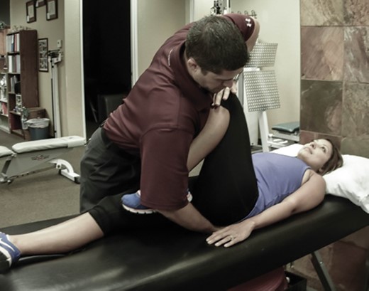
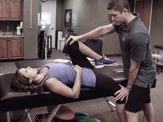
Provocation Tests
A positive provocation test is one that reproduces the patient’s symptoms, suggesting that the joint that has been stressed may be the source of the patient’s pain. Note that a torsional force is applied to both the sacroiliac joint and the hip joint during Patrick’s test, and this test is therefore less able to distinguish between hip and SIJ pain.
SIJ Provocation Tests (Physical Exam Findings)
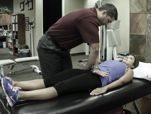
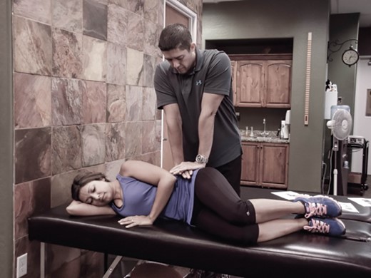
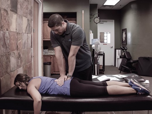
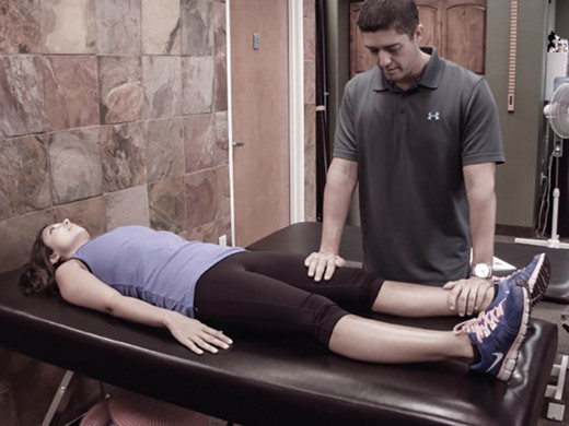
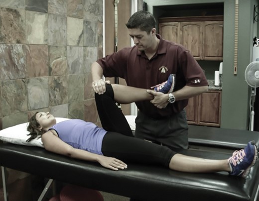
| Test | Description | Photo |
|---|---|---|
| Patrick’s Test |
| 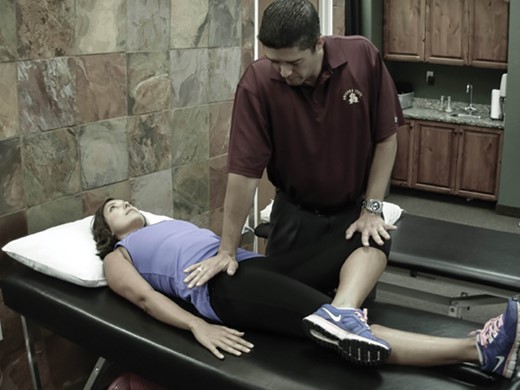 |
| Thigh Thrust |
|  |
| Gaenslen’s Test |
|  |
| Distraction |
|  |
| Compression |
|  |
| Sacral Thrust |
|  |
| Test | Description | Photo |
|---|---|---|
| Patrick’s Test |
| |
| Thigh Thrust |
| |
| Gaenslen’s Test |
| |
| Distraction |
| |
| Compression |
| |
| Sacral Thrust |
| |
ASIS = anterior superior iliac spine; SI = sacroiliac
SIJ Provocation Tests (Physical Exam Findings)
| Test | Description | Photo |
|---|---|---|
| Patrick’s Test |
| |
| Thigh Thrust |
| |
| Gaenslen’s Test |
| |
| Distraction |
| |
| Compression |
| |
| Sacral Thrust |
| |
| Test | Description | Photo |
|---|---|---|
| Patrick’s Test |
| |
| Thigh Thrust |
| |
| Gaenslen’s Test |
| |
| Distraction |
| |
| Compression |
| |
| Sacral Thrust |
| |
ASIS = anterior superior iliac spine; SI = sacroiliac
Hip Provocation Tests (Physical Exam Findings)
| Test | Description | Photo |
|---|---|---|
| Log Roll |
|  |
| Anterior Impingement Test |
|  |
| FABER/Patrick’s Test |
|  |
| Test | Description | Photo |
|---|---|---|
| Log Roll |
| |
| Anterior Impingement Test |
| |
| FABER/Patrick’s Test |
| |
ASIS = anterior superior iliac spine; SI = sacroiliac.
Hip Provocation Tests (Physical Exam Findings)
| Test | Description | Photo |
|---|---|---|
| Log Roll |
| |
| Anterior Impingement Test |
| |
| FABER/Patrick’s Test |
| |
| Test | Description | Photo |
|---|---|---|
| Log Roll |
| |
| Anterior Impingement Test |
| |
| FABER/Patrick’s Test |
| |
ASIS = anterior superior iliac spine; SI = sacroiliac.
References
Author notes
Conflicts of interest: None of the other authors has any financial conflicts of interest to disclose.
