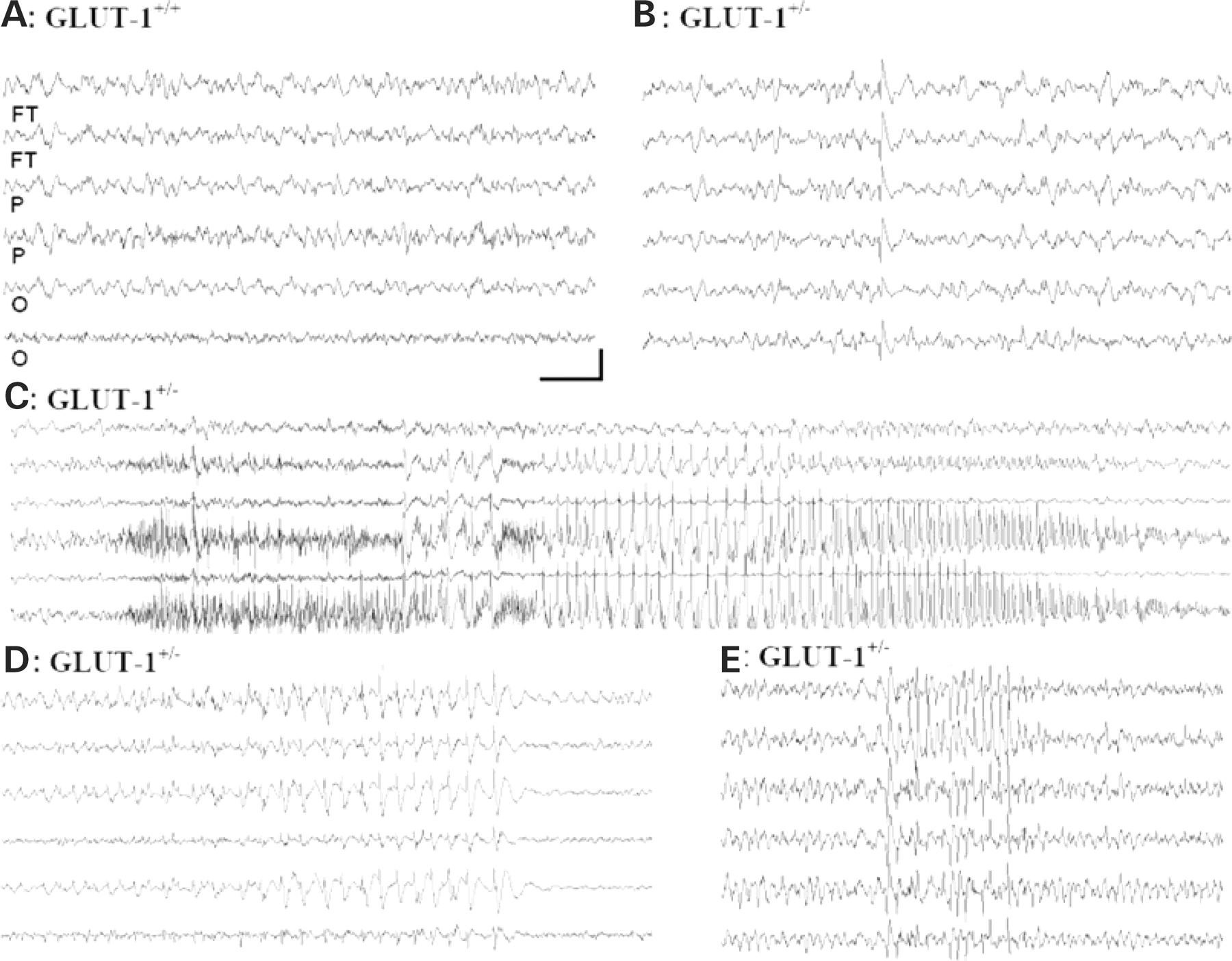
Figure 5. Representative electroencephalographic activity recorded from awake wild-type GLUT-1+/+ (A) and heterozygous GLUT-1+/− mice (B). Traces from left and right hemispheres (alternating from anterior to posterior electrodes). The baseline cortical activity was a relatively normal, low-amplitude desynchronized EEG activity with frequent appearance of solitary generalized interictal discharges with no behavioral correlate. A spontaneous seizure in an adult GLUT-1+/− mouse is shown (C). This electrographic seizure showed a lateralized discharge arising in the right hemisphere with largest amplitude over the parieto-occipital neocortex, and with a decremental EEG pattern over the contralateral hemisphere. Another electrographic seizure pattern, featuring a slow (<3 Hz), high-amplitude, bilateral spontaneous cortical spike-wave synchronous discharge accompanied by normal behavior is shown (D). A spontaneous 6 s−1 rhythmic spiking seizure accompanied by behavioral arrest is shown (E). In all seizures, the EEG activity reverts to normal low-amplitude, high-frequency patterns immediately following the end of the discharge. Calibrations: (A–C, E) 200 mV, 1 s; (D) 200 mV, 1.5 s.