Fig. 5 Localization of OsNHX1–sGFP fusion protein expressed in rice cells. OsNHX1–sGFP fusion protein was expressed in rice cells as described in Materials and Methods. Green fluorescence of sGFP in the cells was imaged under a laser-scanning confocal microscope (A-a and B-a). Tonoplasts are stained red with FM4-64 (B-b). The two images of B-a and B-b are overlaid in B-c. Contrast images of the cells are shown in A-b and B-d. N, nucleus; V, vacuole. Scale bar in A = 50 µm; scale bar in B = 10 µm.
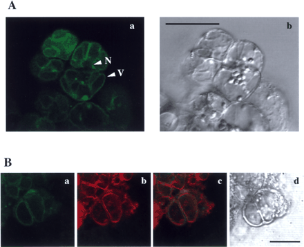
Fig. 5 Localization of OsNHX1–sGFP fusion protein expressed in rice cells. OsNHX1–sGFP fusion protein was expressed in rice cells as described in Materials and Methods. Green fluorescence of sGFP in the cells was imaged under a laser-scanning confocal microscope (A-a and B-a). Tonoplasts are stained red with FM4-64 (B-b). The two images of B-a and B-b are overlaid in B-c. Contrast images of the cells are shown in A-b and B-d. N, nucleus; V, vacuole. Scale bar in A = 50 µm; scale bar in B = 10 µm.

Close
This Feature Is Available To Subscribers Only

Sign In or Create an Account

Close

This PDF is available to Subscribers Only

View Article Abstract & Purchase Options

For full access to this pdf, sign in to an existing account, or purchase an annual subscription.

Close