


A 61-year old patient presenting with an apical pseudoaneurysm after transcatheter aortic valve implantation (Braile Inovare) (Fig. 1). The pseudoaneurysm was punctured and a Foley catheter was placed inside the left ventricle. The Foley catheter was pulled in order to temporarily occlude the pseudoaneurysm (Fig. 2) allowing pledget sutures to close it.
Figure 1:
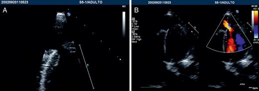
(A) Echocardiogram. An apical view revealing the pseudoaneurysm sac. (B) Echocardiogram. Doppler revealing flow inside the aneurysmal sac.
Figure 2:
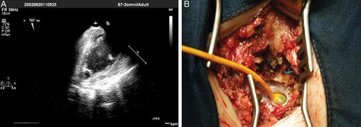
(A) Echocardiogram. A Foley catheter inside the left ventricle cavity. (B) Surgical aspect. Foley catheter in position.
© The Author 2012. Published by Oxford University Press on behalf of the European Association for Cardio-Thoracic Surgery. All rights reserved.
