Figure 2.
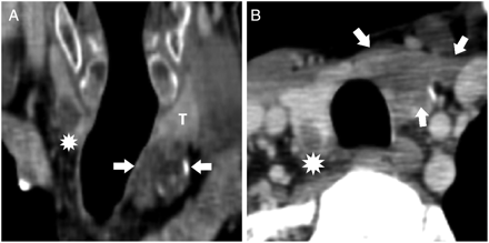
Multisection computed tomography in coronal oblique slices (A) and axial oblique slices (B) showed inhomogeneous, hypodense, partly calcified lesion (arrows) distinctly part of the left thyroid lobe (T) postoperatively confirmed as PC and well-bordered hypodense lesion in posterior part of right thyroid lobe (above asterisk) postoperatively confirmed as metastasis of primary tumor.