Fig. 1.
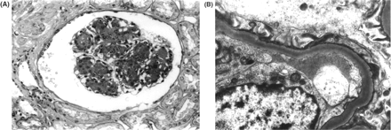
Photomicrographs of biopsies form the patient's native kidney. (A) Light microscopy of a representative glomerulus shows nodules positively stained with periodic acid-Schiff (PAS) in the mesangial area, associated with mesangial hypercellularity (original magnification × 100). (B) Electronmicrograph shows homogenous electron-dense deposits in the glomerular basement membrane (original magnification × 10 000). (C) Immunofluorescence microscopy for λ-light chain displays homogenous deposition in mesangial nodules, and linear deposition along glomerular basement membranes, Bowman's capsule, and tubular basement membranes (original magnification × 200). (D) Paraffin section stained with a polyclonal γ-chain antibody conjugated with peroxidase. The dark-brown reaction product is shown deposited in glomerular and tubular basement membranes (original magnification × 100).
Photomicrographs of biopsies form the patient's native kidney. (A) Light microscopy of a representative glomerulus shows nodules positively stained with periodic acid-Schiff (PAS) in the mesangial area, associated with mesangial hypercellularity (original magnification × 100). (B) Electronmicrograph shows homogenous electron-dense deposits in the glomerular basement membrane (original magnification × 10 000). (C) Immunofluorescence microscopy for λ-light chain displays homogenous deposition in mesangial nodules, and linear deposition along glomerular basement membranes, Bowman's capsule, and tubular basement membranes (original magnification × 200). (D) Paraffin section stained with a polyclonal γ-chain antibody conjugated with peroxidase. The dark-brown reaction product is shown deposited in glomerular and tubular basement membranes (original magnification × 100).

Close
This Feature Is Available To Subscribers Only

Sign In or Create an Account

Close

This PDF is available to Subscribers Only

View Article Abstract & Purchase Options

For full access to this pdf, sign in to an existing account, or purchase an annual subscription.

Close