Figure 1.
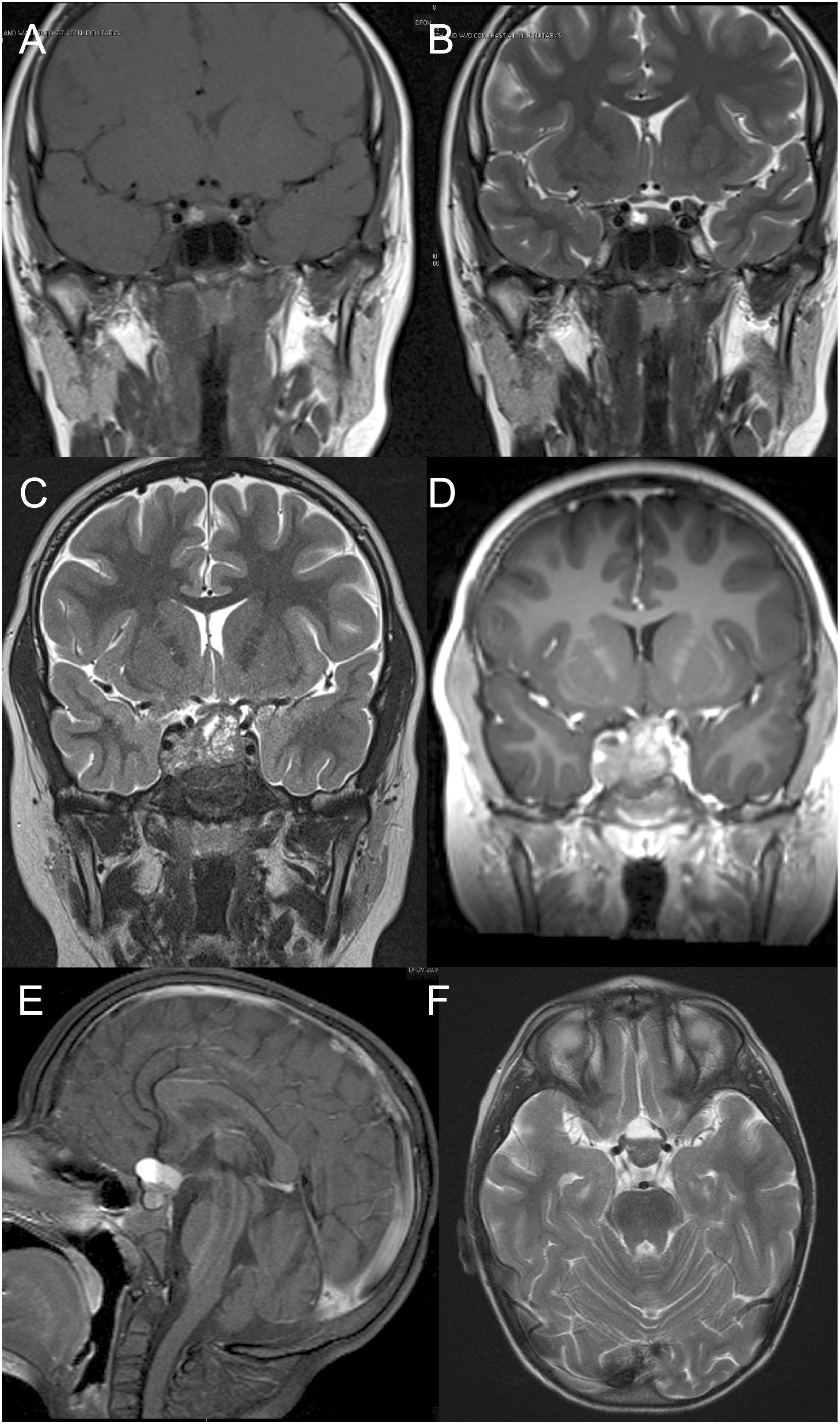
Imaging findings in patients with subclinical pituitary hemorrhage, necrosis, and/or cystic degeneration. A, T1, and B, T2 magnetic resonance imaging (MRI) scans of the same patient showing area of high intensity inside the tumor suggesting acute/subacute episode. C, T2, and D, T1 MRI scans of the same patient showing heterogeneity in a large tumor with high-intensity areas suggesting blood-filled cavities and/or necrosis. E, T1, and F, T2 MRI scans of the same patient showing fluid inside the tumor.