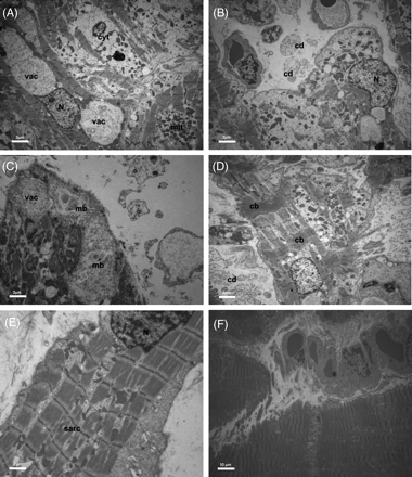
Electron microscopy of ‘acute’ biopsies showing numerous vacuoles of different sizes and contents (myelin bodies, residual cellular products), loss of contractile material, and areas of non-specified cytoplasm (A). The interstitial space was widened containing formation of cellular debris (B). In the ‘acute’ phase, formation of myelin bodies could be documented (C). In TTC contraction bands of sarcomeres were found (D). ‘Recovered’ biopsies showed a nearly complete rearrangement of contractile material with regularly distributed sarcomeres, normal nuclei, and mitochondria (E, F). vac, vacuole; svac, small vacuoles; N, nucleus; cyt, cytoplasm; mit, mitochondria; cd, cellular debris; mb, myelies bodies; sarc, sarcomeres; cb, contraction band.